

Pilomatrixoma
Calcifying epithelioma of Malherbe; pilomatricoma (US spelling)
Pilomatrixoma is a common benign adnexal tumour with differentiation toward the matrical epithelium of the hair bulb, typically presenting as a firm, calcified, dermal-subcutaneous nodule on the head, neck or upper limb of a child or young adult. The clinical hallmark — the "tent sign" / "teeter-totter sign" produced by stretching the overlying skin to reveal multifaceted bony hardness — together with the patient's age, makes the diagnosis straightforward in most cases. Surgical excision is curative. Recognition matters because the lesion is one of the most common benign tumours encountered in paediatric dermatology, plastic surgery and ENT, frequently misdiagnosed as an epidermoid cyst or foreign body. Multiple pilomatrixomas may signal underlying syndromes including myotonic dystrophy, Gardner syndrome, Turner syndrome and Rubinstein-Taybi syndrome. The malignant counterpart — pilomatrix carcinoma — is rare and aggressive.
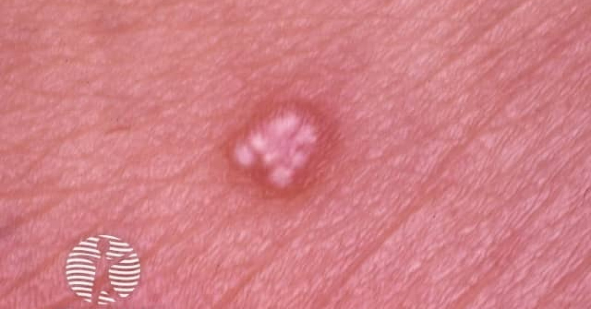
Clinical features
- Solitary, slow-growing, firm-to-hard, often bony-hard dermal-subcutaneous nodule, 0.5–3 cm.
- Skin overlying lesion may be normal, blue-violet, red or stretched-thin and tense.
- "Tent sign" / "teeter-totter sign" — stretching the overlying skin produces multifaceted bony hardness underneath, suggesting the calcified internal structure.
- Distribution — head and neck (~60%), upper extremities, trunk; less often lower limbs.
- Median age 5–15 (most common in children); second peak in 50s–60s.
- Asymptomatic; occasional ulceration / extrusion of calcified material ("perforating pilomatrixoma").
- Multiple pilomatrixomas — consider myotonic dystrophy (commonest association), Gardner syndrome (FAP), Turner syndrome, Rubinstein-Taybi syndrome.
Histology
- Sharply demarcated dermal-subcutaneous nodule with a peripheral rim of basaloid matrical-type cells transitioning into eosinophilic "shadow" (ghost) cells with lost nuclei but preserved cellular outlines — pathognomonic of matrical differentiation.
- Calcification (60–75%) and ossification within the lesion.
- Foreign-body giant cell reaction often present at the periphery.
- No atypia, mitoses or necrosis in benign pilomatrixoma.
- Immunohistochemistry — strong nuclear β-catenin accumulation reflecting CTNNB1 mutation.
- Differential by histology: pilomatrix carcinoma (atypia, mitoses, necrosis, infiltrative growth — see monograph); matrical SCC; foreign-body granuloma.
Multiple pilomatrixomas — associated syndromes
- Myotonic dystrophy (DM1) — commonest association of multiple pilomatrixomas; suspect in any patient with three or more pilomatrixomas, particularly if family history.
- Gardner syndrome (FAP variant) — pilomatrixomas may co-exist with epidermoid cysts, fibromas, desmoid tumours and gastrointestinal polyposis.
- Turner syndrome (45,X).
- Rubinstein-Taybi syndrome.
- Sotos syndrome, Kabuki syndrome — case reports.
- Always ask about family history and other features when encountering multiple pilomatrixomas.
Management
- Surgical excision with narrow (3–5 mm) margins is curative; histology is mandatory.
- Recurrence after complete excision <3%.
- Imaging not required for typical solitary lesions; ultrasound shows characteristic well-circumscribed hyperechoic nodule with internal calcification and posterior shadowing — useful when clinical diagnosis uncertain.
- If histology shows atypical or carcinomatous features — manage as pilomatrix carcinoma.
- If multiple pilomatrixomas — clinical evaluation for myotonic dystrophy / Gardner / Turner / Rubinstein-Taybi; refer to clinical genetics where appropriate.
References
- Lan MY et al. Pilomatricoma of the head and neck — a retrospective review of 179 cases. Arch Otolaryngol Head Neck Surg; 2003.
- Chan EF et al. A common skin tumour is caused by activating mutations in beta-catenin. Nat Genet; 1999.
Spot a correction?
If any clinical statement, citation or link on this page needs updating, please email admin@skinoncology.net with the page name, the proposed correction and the supporting source.