

Epidermoid cyst
Epidermal inclusion cyst; "sebaceous cyst" (a misnomer — these are not derived from sebaceous glands); follicular infundibular cyst
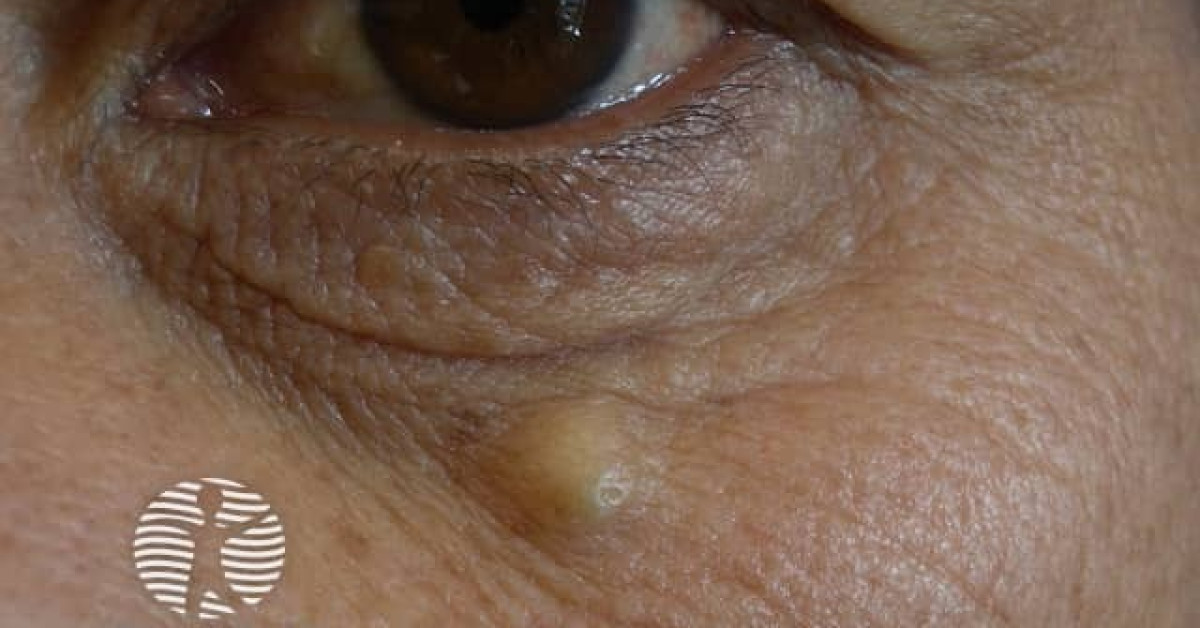
The epidermoid cyst is the commonest cutaneous cyst, presenting as a slow-growing, mobile, dome-shaped subcutaneous nodule with a characteristic central punctum (representing the follicular infundibulum from which the cyst arises) and, on incision, expressed cheesy malodorous keratin. It is the lesion most often referred to as a "sebaceous cyst" — a misnomer because the cyst contains keratin and is lined by stratified squamous epithelium identical to that of the follicular infundibulum, not sebaceous gland material. Recognition has skin-oncology relevance because: (1) multiple epidermoid cysts in unusual sites (face, scalp, back, often appearing in childhood) are an early marker of Gardner syndrome, the FAP variant; (2) proliferating trichilemmal cysts and rare malignant transformation in long-standing cysts can mimic SCC; and (3) cysts are frequent BCC / soft-tissue tumour clinical mimics.
Clinical features
- Slow-growing, mobile, dome-shaped subcutaneous nodule, 0.5–5 cm.
- Central punctum representing the follicular infundibulum — the diagnostic clue.
- Distribution — face, neck, trunk (especially upper back, chest), scrotum; less often extremities.
- Asymptomatic until traumatised, infected or spontaneously ruptured.
- Rupture into the dermis triggers a foreign-body inflammatory reaction — painful red swollen "infected cyst" appearance, but usually sterile.
- Multiple cysts in childhood or unusual locations — consider Gardner syndrome.
Histology
- Cyst lined by stratified squamous epithelium with a granular layer (identical to follicular infundibular epithelium and surface epidermis), surrounded by collagenous stroma.
- Cyst cavity filled with concentric layers of orthokeratotic keratin ("lamellar keratin").
- No atypia in benign epidermoid cyst.
- If ruptured — foreign-body giant-cell reaction, granulomatous inflammation, sometimes pseudoepitheliomatous hyperplasia (mimicking SCC).
- Variants:
- Pilar cyst (trichilemmal cyst) — see monograph; lining without granular layer; abrupt keratinisation; commoner on scalp.
- Steatocystoma — sebaceous-gland-lined cyst; multiple in steatocystoma multiplex.
- Hybrid cyst — features of more than one type.
Multiple cysts → Gardner syndrome
- Multiple epidermoid cysts in childhood or in unusual sites (face, scalp, back) should prompt consideration of Gardner syndrome — an autosomal dominant variant of familial adenomatous polyposis (APC mutation).
- Other Gardner features: osteomas (skull, mandible), desmoid tumours, fibromatosis, pilomatrixomas, congenital hypertrophy of retinal pigment epithelium (CHRPE), supernumerary teeth, gastrointestinal polyposis with near-100% lifetime colorectal cancer risk.
- Refer to clinical genetics for germline APC testing and to gastroenterology for colonoscopic surveillance.
Malignant transformation — rare but reported
- Squamous cell carcinoma arising in a long-standing epidermoid cyst — exceptionally rare but reported.
- Warning signs:
- Abrupt growth in a previously stable cyst.
- Ulceration.
- Bleeding.
- Fixity to deeper tissues.
- Pain disproportionate to the lesion.
- Any change should prompt excision with full histology.
- Pseudoepitheliomatous hyperplasia in a ruptured cyst can be misdiagnosed as SCC — request histopathology by an experienced dermatopathologist.
Management
- Asymptomatic non-inflamed cysts — no treatment necessary; reassurance.
- Symptomatic, inflamed, infected or unsightly cysts — surgical excision with the cyst wall intact (incomplete removal of the wall produces recurrence).
- Acutely inflamed cysts — incise and drain plus oral flucloxacillin / clindamycin; arrange for elective excision once inflammation has settled (6–8 weeks).
- Multiple cysts in childhood / unusual sites — clinical genetics referral for Gardner syndrome workup.
- Histology of any unusual / atypical / changing cyst.
References
- Hoang VT et al. Epidermoid cyst — review of pathology and management. J Cutan Med Surg; 2019.
- Brown N et al. Cutaneous manifestations of Gardner syndrome. Dermatol Online J; 2018.
Spot a correction?
If any clinical statement, citation or link on this page needs updating, please email admin@skinoncology.net with the page name, the proposed correction and the supporting source.