Naevus lipomatosus superficialis
Naevus lipomatosus cutaneous superficialis · NLCS · superficial lipomatous naevus · fat naevus · Hoffmann-Zurhelle naevus
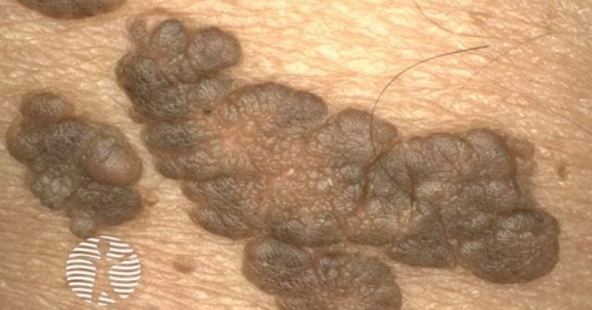
Naevus lipomatosus superficialis is a rare benign cutaneous hamartoma in which mature adipocytes are present ectopically in the dermis without continuity with subcutaneous fat. The classical Hoffmann-Zurhelle type presents at birth or in the first three decades as grouped, soft, skin-coloured or yellowish papules/nodules/plaques, usually on the lower back, buttock or upper thigh. A solitary adult type presents as a single soft papule or subcutaneous swelling anywhere. The main role for skin-oncology clinicians is diagnosis, reassurance, and excision only for symptoms, ulceration, appearance or uncertainty.
Clinical types
- Classical type: present at birth or appears in the first three decades; grouped soft papules, nodules or plaques, often on lower back, buttock, pelvic girdle or upper thigh.
- Classical lesions may be linear, segmental or zosteriform; the surface can be smooth, wrinkled, pedunculated, sessile, cerebriform, hairy or comedo-like.
- Solitary type: tends to arise after the second decade as a single soft, yellow or skin-coloured dome-shaped papule, nodule or swelling anywhere on the body.
- Usually asymptomatic; the main concern is appearance or mechanical irritation.
- Rarely, large lesions can ulcerate or undergo superficial necrosis from trauma or pressure.
Histology and diagnosis
- Diagnosis is confirmed histologically.
- Mature adipocytes are found in the dermis, often between collagen bundles and around vessels or adnexal structures.
- The adipocytes are not connected to the subcutaneous fat, which helps distinguish the lesion from ordinary lipoma.
- The overlying epidermis may show papillomatosis, acanthosis or basal pigmentation.
- Biopsy or excision is reasonable if the lesion is atypical, ulcerated, enlarging, diagnostically uncertain or clinically mimics another tumour.
Differential diagnosis
- Classical type: neurofibromatosis, lipoma, sebaceous naevus, verrucous epidermal naevus and connective-tissue naevus.
- Solitary type: acrochordon/fibroepithelial polyp, lipoma, accessory nipple, lymphatic malformation, haemangioma, neurofibroma, trichoepithelioma and cylindroma.
- Soft cerebriform plaques on the lower trunk/buttock should also prompt consideration of plexiform neurofibroma and focal dermal hypoplasia if syndromic features are present.
- If a presumed skin tag or lipoma has unusual texture, congenital history, grouped distribution or marked dermal component, histology is helpful.
Management
- No treatment is required for a typical, asymptomatic lesion once diagnosis is secure.
- Excision is the preferred treatment when intervention is needed for cosmesis, repeated trauma, ulceration, symptoms or diagnostic uncertainty.
- Large classical lesions may need staged excision or reconstructive planning if removal is requested.
- Laser/ablative approaches have been reported but may leave residual dermal adipocytes and are less definitive than excision.
- There is no recognised malignant transformation risk; prognosis is excellent.
Clinical pitfalls
- Do not assume every soft pedunculated lesion is a skin tag; congenital grouped or cerebriform lesions deserve a wider differential.
- Do not diagnose ordinary lipoma if histology shows adipocytes within dermis rather than a subcutaneous encapsulated fat tumour.
- Ulceration is uncommon and should trigger review for trauma, pressure and alternative diagnoses.
- Document extent before treatment; removal for appearance can be reconstructively more involved than the benign diagnosis suggests.
References
- DermNet. Naevus lipomatosus superficialis. Updated January 2020.
- Lima CDS et al. Nevus lipomatosus cutaneous superficialis. An Bras Dermatol. 2017;92:711-713.
- Goucha S et al. Nevus lipomatosus cutaneous superficialis: report of eight cases. Dermatol Ther (Heidelb). 2011;1:25-30.
- Yap FB. Nevus lipomatosus superficialis. Singapore Med J. 2009;50:e161-e162.
Spot a correction?
If any clinical statement, citation or link on this page needs updating, please email admin@skinoncology.net with the page name, the proposed correction and the supporting source.