

Lymphangioma circumscriptum
Microcystic lymphatic malformation; localised lymphatic malformation; lymphangioma circumscriptum
Lymphangioma circumscriptum is a microcystic lymphatic malformation presenting as a localised group of small, fluid-filled vesicles resembling frog spawn on the skin, often with overlying haemorrhagic foci giving a black-red papular appearance. Most are congenital or present in infancy / early childhood, though adult-onset cases occur (often post-traumatic or post-RT). Sites — axilla, proximal limbs, neck, oral cavity, vulva, scrotum. The lesion is benign but problematic for lymph leakage, recurrent cellulitis and cosmetic / psychosocial impact. Treatment is challenging — surgical excision with wide deep margins, sclerotherapy (OK-432, doxycycline), CO₂ laser; recurrence is the rule rather than the exception.
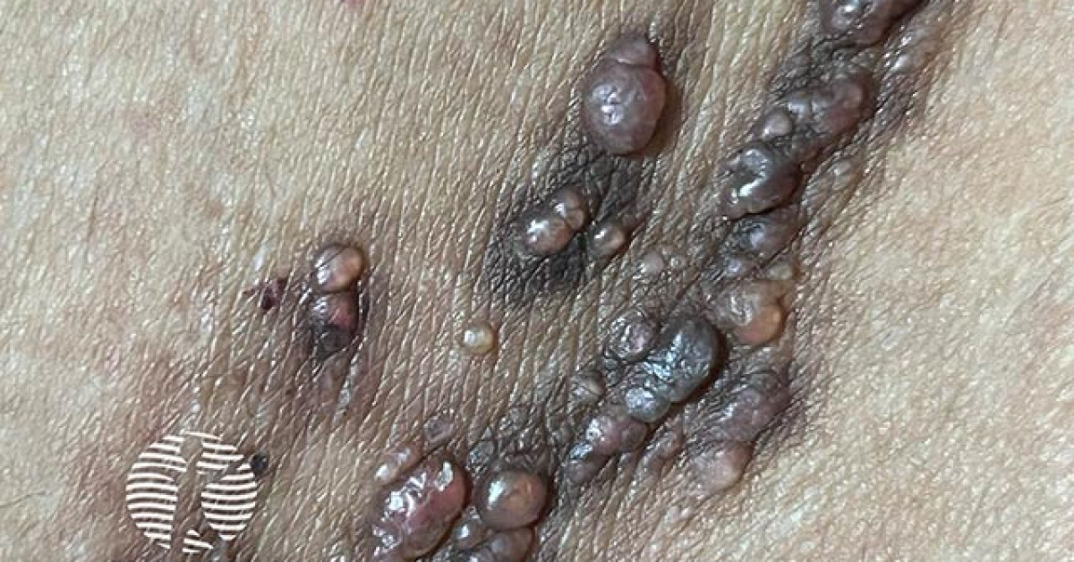
Clinical features
- Localised group of clear or haemorrhagic 1–4 mm vesicles ("frog spawn"); occasionally larger fluid-filled cysts.
- Surface can be verrucous in long-standing lesions.
- Spontaneous leakage of clear lymph fluid; occasional bleeding.
- Common sites — axilla, proximal upper limb, neck, oral cavity, vulva, scrotum.
- Onset — congenital or early childhood (most); secondary lymphangioma may follow trauma, surgery, RT or chronic lymphoedema.
- Recurrent cellulitis is a recognised complication.
Differential
- Herpetic lesions — vesicles, painful, evolving over days.
- Hidradenitis suppurativa with sinus tracts — axillary distribution overlap.
- Verrucous epidermal naevus.
- Mucinous metaplasia, angiokeratoma circumscriptum.
- Pyogenic granuloma if haemorrhagic vesicles predominate.
- Acquired lymphangiectasia — usually post-lymphoedema or post-RT; clinically similar to LC.
Histology
- Dilated thin-walled lymphatic vessels in the papillary dermis lined by D2-40-positive (podoplanin-positive) endothelium.
- Vessels often communicate with deeper, larger cisterns of fluid in the reticular dermis and subcutis — the explanation for high recurrence after superficial removal.
- Microscopic extension well beyond clinical margins is the rule.
Management
- No treatment is curative; recurrence is common.
- Conservative — observation, compression, antibiotic prophylaxis for recurrent cellulitis.
- Surgical excision with wide deep margins (to fascia) — best chance of long-term control but recurrence occurs from deeper cisterns; reserve for symptomatic / cosmetic indication.
- Sclerotherapy — OK-432 (picibanil), doxycycline, sodium tetradecyl sulphate; better for macrocystic components.
- CO₂ laser ablation, electrocautery — superficial; high recurrence.
- Radiotherapy — historic; rarely used now due to long-term cancer risk.
- Sirolimus — emerging systemic option for complex lymphatic anomalies.
- Specialist vascular-anomaly MDT input.
References
- Whimster IW. The pathology of lymphangioma circumscriptum. Br J Dermatol; 1976.
- Adams MT et al. Sirolimus for complicated lymphatic malformations. Pediatrics; 2016.
Spot a correction?
If any clinical statement, citation or link on this page needs updating, please email admin@skinoncology.net with the page name, the proposed correction and the supporting source.