Epidermal naevus
Keratinocytic epidermal naevus · verrucous epidermal naevus · linear epidermal naevus · epidermal nevus
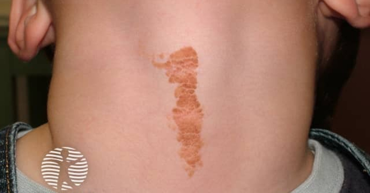
An epidermal naevus is a benign hamartomatous overgrowth of epidermis, usually present at birth or arising in early childhood. Linear lesions follow Blaschko lines and often start as flat tan-brown marks before becoming thickened, papillomatous or warty. Most are isolated and benign. The clinical task is to recognise persistent Blaschko-linear epidermal lesions, separate them from inflammatory mimics such as lichen striatus and linear psoriasis, and identify extensive/systematised lesions or extracutaneous features that suggest an epidermal naevus syndrome.
Biology and classification
- Most lesions are keratinocytic epidermal naevi: hamartomatous overgrowth of keratinocytes.
- Post-zygotic mosaicism explains the Blaschko-linear distribution; reported genes include FGFR3, PIK3CA and HRAS, depending on phenotype.
- Recognised patterns include linear epidermal naevus, systematised epidermal naevus, epidermolytic epidermal naevus, acantholytic epidermal naevus and linear porokeratosis-like lesions.
- When appendageal structures predominate, the term organoid naevus is used, including sebaceous naevus and naevus comedonicus.
- ILVEN is often considered separately: pruritic, erythematous and inflammatory, usually along Blaschko lines.
Clinical features
- About half are present at birth; many others appear during infancy or early childhood.
- Usually unilateral, linear or whorled along Blaschko lines on trunk or limbs; face/scalp involvement is less common for ordinary keratinocytic lesions.
- Early lesions may be flat tan or brown macules/patches; with age they become thicker, papillomatous, verrucous or hyperkeratotic.
- May enlarge proportionately with growth and can become more conspicuous around puberty.
- Most are asymptomatic; itch, erythema and psoriasiform inflammation suggest ILVEN or an inflammatory mimic.
Syndrome red flags
- Extensive, systematised, bilateral or very large lesions need a broader review for extracutaneous abnormalities.
- Epidermal naevus syndromes can involve the central nervous system, eyes and skeleton; examples include Schimmelpenning syndrome, naevus comedonicus syndrome, CHILD syndrome and Proteus/CLOVES-spectrum disorders.
- Ask about seizures, developmental delay, headaches, visual symptoms, limb asymmetry, scoliosis, bony overgrowth and recurrent infections.
- Consider paediatric dermatology/genetics referral when lesions are extensive, syndromic features are present, or the diagnosis overlaps with vascular/overgrowth syndromes.
Differential diagnosis
- Lichen striatus: usually acquired, inflammatory, self-limiting and more common in children.
- Linear lichen planus, linear psoriasis or linear porokeratosis.
- ILVEN: intensely pruritic, persistent, inflammatory and often treatment-resistant.
- Incontinentia pigmenti: staged vesicular, verrucous and pigmentary evolution, typically in female infants.
- Naevus sebaceus, naevus comedonicus, Becker naevus and connective-tissue naevi.
- Verruca vulgaris, especially if a localised verrucous lesion appears later in life.
Management
- Reassure for small, stable, asymptomatic lesions.
- Emollients and keratolytics can help scale/roughness; topical calcipotriol or topical retinoids may reduce thickness in selected cases but responses are variable.
- Topical corticosteroids/calcineurin inhibitors are useful only for inflammatory components, not for the naevus itself.
- Laser ablation, shave/dermabrasion or excision can be considered for symptoms, repeated trauma, diagnostic uncertainty or appearance, but counsel about scarring and recurrence.
- Biopsy any new nodular, ulcerated, bleeding, rapidly changing or atypical focus within a longstanding lesion.
References
- DermNet. Epidermal naevi.
- MedlinePlus Genetics. Epidermal nevus.
- NCBI Bookshelf. Epidermal Nevus Syndromes.
- Groesser L et al. Postzygotic HRAS and KRAS mutations cause naevus sebaceous and Schimmelpenning syndrome. Nat Genet. 2012;44:783-787.
Spot a correction?
If any clinical statement, citation or link on this page needs updating, please email admin@skinoncology.net with the page name, the proposed correction and the supporting source.