

Ink-spot lentigo
reticulated black solar lentigo · reticulated lentigo · solar reticulated lentigo · ink-spot reticulated lentigo
Ink-spot lentigo is a benign solar lentigo variant with a characteristic dramatically dark, jagged, reticulated pattern that mimics melanoma in situ — particularly lentigo maligna — on chronically sun-damaged skin of older fair-skinned adults. Despite its alarming appearance, dermoscopy shows a sharply circumscribed, irregular, dark pigment network without melanoma-specific features. Recognition is essential to avoid unnecessary excision, while ensuring true LM is not missed.
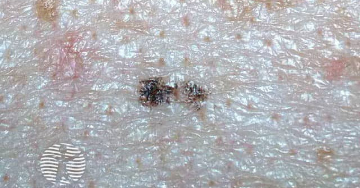
Clinical features
- Single or few sharply demarcated dark brown / black macules.
- Irregular, jagged, "ink-blot"-like outline.
- 1-5 mm diameter typically; can be larger.
- Sites: shoulders, upper back, dorsal forearms — chronically sun-damaged skin.
- Older adults; fair Fitzpatrick I-II skin; high lifetime UV exposure.
- Stable; slow growth at most.
- Often co-exists with conventional solar lentigines.
Dermoscopy
- Sharply circumscribed dark pigment network — the hallmark.
- Network composed of regular thick interconnecting lines and small holes.
- Symmetric structure within the irregular outline.
- No melanoma features:
- No atypical pigment network.
- No blue-white veil.
- No regression / shiny white structures.
- No pseudopods, streaks, or atypical globules.
- No asymmetric pigmented follicular openings (LM features).
- Sometimes "fingerprint" pattern reminiscent of solar lentigo.
Differentials
- Lentigo maligna — pseudo-network with asymmetric pigmented follicular openings; rhomboidal structures; slate-grey dots; biopsy if uncertain.
- Pigmented actinic keratosis — keratotic scale; pseudo-network.
- Solar lentigo (typical) — moth-eaten edge; uniform light tan pigment.
- Atypical naevus / dysplastic naevus.
- Pigmented BCC — pearly border, blue-grey ovoid nests, arborising vessels.
- Naevoid melanoma — biopsy if any atypia.
- Pigmented seborrhoeic keratosis — milia-like cysts.
Management
- Reassurance — benign solar lentigo variant.
- If diagnostic uncertainty at dermoscopy / clinical examination → excisional biopsy (preferred to shave / punch) to exclude lentigo maligna.
- Cosmetic management:
- Q-switched / picosecond laser for pigment.
- Cryotherapy (caution about hypopigmentation in Fitzpatrick III-VI).
- Tretinoin / hydroquinone (modest).
- Counsel about photoprotection given background of chronic UV damage.
- Document with photography; surveillance if multiple atypical-pigmented lesions on background of solar damage.
- Consider field-cancerisation assessment — look for actinic keratoses, Bowen disease, BCC.
References
- Bolognia JL. Reticulated black solar lentigo ("ink spot" lentigo). Arch Dermatol. 1992;128:934-940.
- Carrera C et al. Dermoscopy of facial lentigines: ink-spot lentigo / reticulated lentigo. Br J Dermatol. 2018;179:1199-1200.
- Stolz W, Argenziano G. Dermoscopy of pigmented skin lesions. Atlas of Dermoscopy; 2002.
- British Association of Dermatologists. Lentigines — patient information leaflet. London: BAD; 2023.
Spot a correction?
If any clinical statement, citation or link on this page needs updating, please email admin@skinoncology.net with the page name, the proposed correction and the supporting source.