Lentigo simplex
Simple lentigo; juvenile lentigo; lentigines simplex
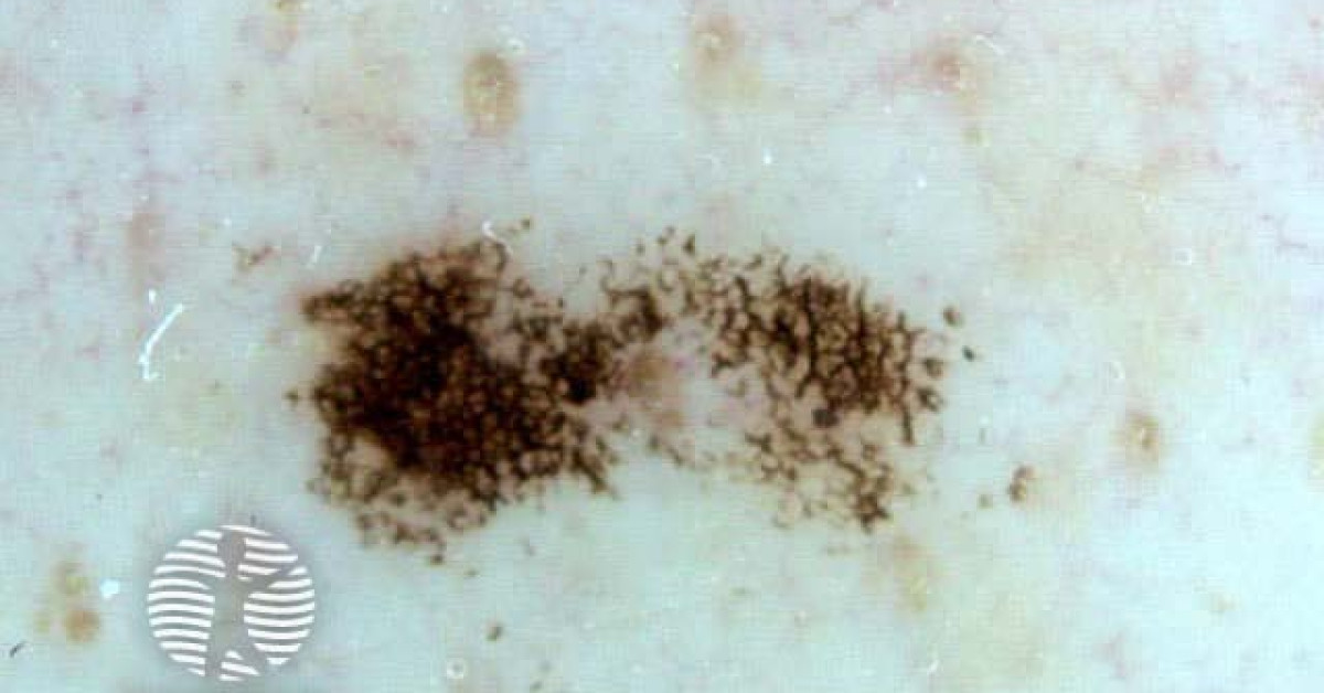
Lentigo simplex is the most fundamental pigmented melanocytic macule — a small (1–5 mm), well-defined, uniformly pigmented brown macule that develops in childhood and young adulthood, independent of UV exposure. Histologically it is characterised by hyperpigmentation of basal keratinocytes with a modest increase in melanocyte density along an elongated rete ridge (without the irregular junctional nests of a junctional naevus). The relevance in skin oncology is largely as a differential for the more concerning pigmented entities — solar lentigo, ephelis, junctional melanocytic naevus and early lentigo maligna — and as a feature of several genetic syndromes (Peutz-Jeghers, LEOPARD, Carney complex).
Clinical features
- Small (1–5 mm), well-circumscribed, uniformly pigmented brown to dark-brown macule.
- Onset childhood / young adulthood; persists into adult life.
- UV-independent in onset (contrasts with solar lentigo).
- Sites — anywhere on the body; mucosal lentigines (lip vermilion, oral, genital) are well-recognised.
- Usually a single or few lesions; multiple lentigines should prompt consideration of a syndromic context.
Differential
- Ephelis (freckle) — small (1–3 mm), tan; appears in childhood; UV-dependent (darkens with sun, fades in winter); flat, irregular border.
- Solar lentigo — larger (often > 5 mm), older patient, chronically sun-damaged skin (face, dorsum of hands), uniform tan-brown.
- Junctional melanocytic naevus — slightly larger, often darker, with characteristic dermoscopic network.
- Lentigo maligna — older patient, larger, irregular border, multiple shades; biopsy any new or changing pigmented macule on chronically sun-damaged skin.
- Café-au-lait macule — light tan, larger, often single uniform colour.
- Becker naevus — large hyperpigmented patch with hypertrichosis.
Syndromic lentigines
- Peutz-Jeghers syndrome — mucocutaneous lentigines on lips, oral mucosa, periorificial skin + GI hamartomatous polyposis; STK11 mutation; cancer surveillance per UK Lynch-equivalent protocols.
- LEOPARD syndrome / Noonan with multiple lentigines — RASopathy; multiple lentigines + cardiac conduction abnormalities + ocular hypertelorism + pulmonary stenosis + sensorineural deafness.
- Carney complex — spotty mucocutaneous pigmentation + cardiac myxomas + endocrine tumours + blue naevi.
- Laugier-Hunziker syndrome — benign acquired pigmentation of lips, buccal mucosa and nails (longitudinal melanonychia); no malignancy risk.
- Lentiginosis with associations — Bannayan-Riley-Ruvalcaba (PTEN), Cronkhite-Canada.
Management
- Reassurance — no treatment required for typical solitary lentigo simplex.
- Cosmetic — Q-switched laser, IPL, fractional ablative laser; cryotherapy.
- Biopsy any pigmented macule with atypical clinical or dermoscopic features.
- Multiple lentigines, particularly mucosal — consider syndromic context and refer to clinical genetics if appropriate.
- Counselling — photoprotection, self-examination.
References
- Mihm MC Jr, Clark WH Jr. The lentigo as a clinicopathologic entity. Hum Pathol; 1973.
Spot a correction?
If any clinical statement, citation or link on this page needs updating, please email admin@skinoncology.net with the page name, the proposed correction and the supporting source.