

Fibrous papule of the face (facial angiofibroma)
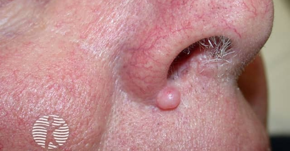
Solitary facial angiofibroma; fibrous papule of the nose
Fibrous papule of the face is one of the commonest benign facial papules in adults — a solitary, firm, dome-shaped, skin-coloured to faintly pink papule, almost invariably on the nose, that represents a benign angiofibroma (dermal fibroblastic proliferation with dilated vessels). Despite its frequency it is a regular diagnostic mimic of small BCC, intradermal melanocytic naevus and inflamed cyst. Multiple early-onset angiofibromas of the face are a major criterion of tuberous sclerosis and should not be diagnosed as solitary fibrous papules.
Clinical features
- Solitary, firm, dome-shaped, skin-coloured to pink papule, 2–5 mm.
- Almost always on the nose (tip, ala, alar crease); occasionally upper lip, chin or forehead.
- Slow growth; long history of stability is typical.
- Median age 30–60; both sexes.
- Asymptomatic.
Dermoscopy
- White structureless background with fine, regular telangiectasias and a subtle whitish halo.
- No pearly margin, no blue-grey nests, no leaf-like structures — features that argue against BCC.
- Useful adjunct but does not replace biopsy where uncertainty persists.
Histology
- Dermal proliferation of stellate and spindled fibroblasts within a fibrotic stroma.
- Dilated thin-walled vessels in the upper dermis.
- Overlying epidermis is generally unremarkable or mildly atrophic.
- Histology is identical to the multiple angiofibromas seen in tuberous sclerosis — the distinction is clinical (solitary vs multiple, onset age).
Differential
- BCC — pearly, telangiectatic, growth over months to years, may ulcerate. Biopsy if any doubt.
- Intradermal melanocytic naevus — softer, often skin-coloured; long stability.
- Trichoepithelioma — typically multiple in Brooke-Spiegler.
- Sebaceous hyperplasia — yellow lobules with crown vessels.
- Inflamed epidermoid cyst.
- Adenoma sebaceum (multiple angiofibromas) of tuberous sclerosis — multiple lesions from childhood/adolescence.
Management
- Confident clinical diagnosis with long stability — reassurance; no treatment required.
- Diagnostic uncertainty — shave or punch excision for histology. Excision biopsy preferred if BCC suspected.
- Cosmetic treatment — shave excision, light electrodesiccation, CO₂ or pulsed-dye laser; counsel about small scar.
- Multiple lesions, particularly early-onset — refer for tuberous sclerosis assessment.
References
- Ackerman AB, Viragh PA. Fibrous papule of the face — a misnomer. Am J Dermatopathol; 1980.
- McGavran MH, Binnington B. Solitary angiofibroma — clinicopathologic study. Arch Dermatol; 1966.
Spot a correction?
If any clinical statement, citation or link on this page needs updating, please email admin@skinoncology.net with the page name, the proposed correction and the supporting source.