Pilar (trichilemmal) cyst
Trichilemmal cyst; isthmus-catagen cyst; scalp cyst; "wen" (older); proliferating trichilemmal cyst (PTC) — distinct neoplastic variant with malignant potential
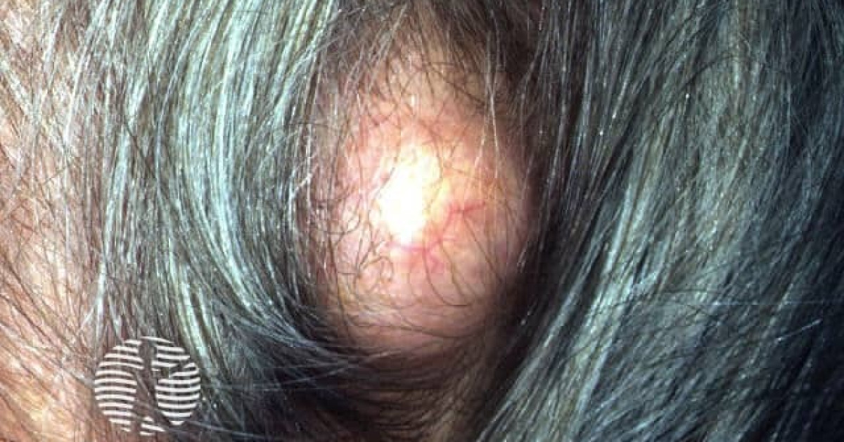
Pilar (trichilemmal) cysts are common, autosomal dominantly inherited cutaneous cysts derived from the outer root sheath of the hair follicle. They classically occur on the scalp of middle-aged adults, where they form smooth, mobile, often multiple subcutaneous nodules without a central punctum (distinguishing them clinically from epidermoid cysts). The condition is benign, but the spectrum extends to proliferating trichilemmal cyst (PTC) — a distinct low-grade neoplastic variant most common on the scalp of elderly women, which can recur and rarely transform to malignant proliferating trichilemmal tumour with regional and distant metastatic potential. Multiple pilar cysts in a young patient may signal a familial autosomal dominant predisposition.
Clinical features
- Smooth, mobile, dome-shaped subcutaneous nodule, 0.5–5 cm, without a central punctum (a distinguishing feature from epidermoid cyst).
- Distribution — >90% on the scalp; less often face, neck, trunk, scrotum.
- Often multiple — autosomal dominant inheritance; positive family history common.
- Median age — adults; F>M.
- Slow-growing; asymptomatic until traumatised, infected, or large enough to compress on a hat.
- Spontaneous rupture is much rarer than for epidermoid cysts.
Histology & spectrum
- Pilar cyst:
- Cyst lined by stratified squamous epithelium without a granular layer — the histological hallmark distinguishing it from epidermoid cyst.
- Abrupt keratinisation with eosinophilic compact keratin filling the cavity.
- Frequent calcification.
- Proliferating trichilemmal cyst (PTC):
- Lobulated, multinodular, partly cystic and partly solid lesion.
- Trichilemmal-type keratinisation; cytological atypia and mitoses present in higher-grade lesions.
- Pushing rather than infiltrative borders in benign / low-grade PTC.
- Recurrence common; rare distant metastasis.
- Malignant proliferating trichilemmal tumour (formerly "malignant proliferating trichilemmal cyst" / "trichilemmal carcinoma in some classifications"):
- Marked atypia, infiltrative deep border, lymphovascular and perineural invasion, tumour necrosis.
- Behaves as a carcinoma with regional and distant metastatic potential.
- Manage as soft-tissue malignancy through skin cancer / sarcoma MDT.
Management
- Asymptomatic pilar cysts — no treatment required; reassurance.
- Symptomatic / large / cosmetically intrusive lesions — surgical excision with intact wall (recurrence if wall left behind).
- Multiple cysts — staged excision; minimally invasive techniques (small incision, "minimal-excision" punch) for cosmesis.
- Suspected proliferating trichilemmal cyst (firm, growing, ulcerated, bleeding) — wide local excision with at least 1 cm margins, full histology.
- Histological diagnosis of malignant proliferating trichilemmal tumour — refer to skin cancer / sarcoma MDT; wide local excision; consider sentinel lymph node biopsy and adjuvant radiotherapy.
References
- Folpe AL et al. Proliferating trichilemmal tumors — clinicopathologic study. J Am Acad Dermatol; 2003.
- Satyaprakash AK et al. Proliferating trichilemmal tumors — review. Dermatol Surg; 2007.
Spot a correction?
If any clinical statement, citation or link on this page needs updating, please email admin@skinoncology.net with the page name, the proposed correction and the supporting source.