

Cutaneous horn
Cornu cutaneum; "human horn"
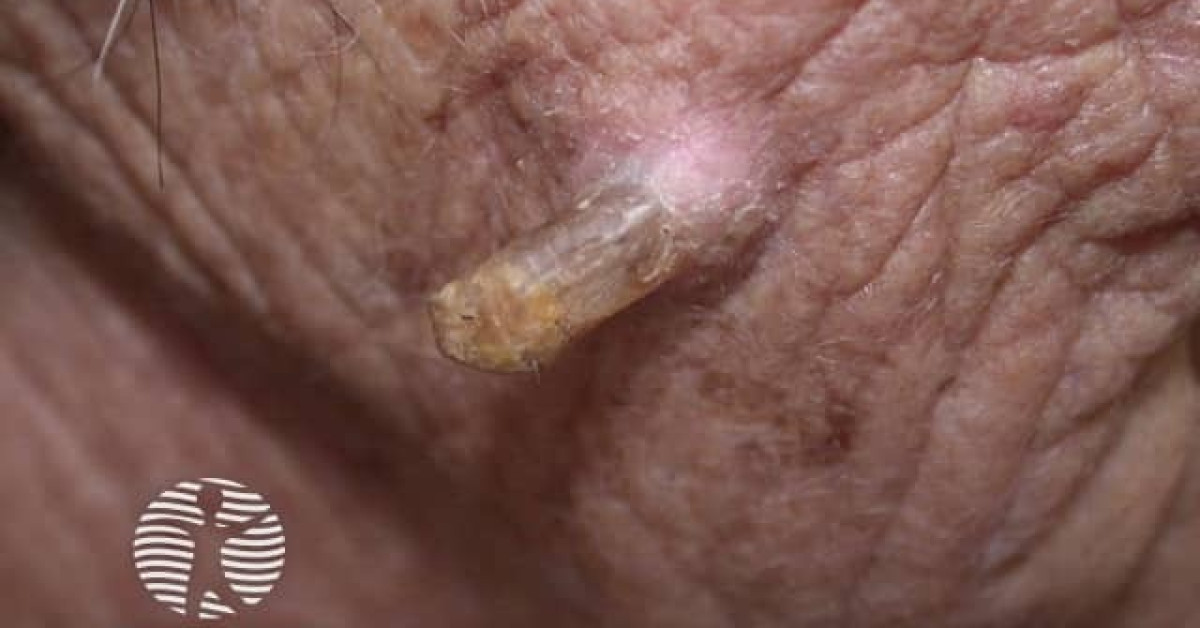
A cutaneous horn (cornu cutaneum) is a clinical descriptive term for a discrete, firm, conical hyperkeratotic projection of compacted keratin growing from the skin — historically the source of "human horn" curiosities in medieval medical writing. Critically, it is NOT a histological diagnosis: any cutaneous horn must be excised with the underlying skin and submitted for histology, because the underlying base will reveal a benign, pre-malignant or malignant lesion in approximately 60% / 23% / 17% of cases respectively. The most common malignant base is squamous cell carcinoma; less commonly basal cell carcinoma, keratoacanthoma, sebaceous carcinoma, melanoma and Merkel cell carcinoma. Risk factors for malignancy at the base include older age, larger diameter, broader base, tenderness, induration and a sun-exposed site. Shave biopsy of the horn alone is inadequate — the diagnosis depends entirely on the underlying skin.
Clinical features
- Conical, firm, yellow-brown to white hyperkeratotic projection rising from the skin surface, ranging from a few millimetres to several centimetres in length.
- Distribution — sun-exposed sites (face, scalp, ears, dorsal hands, forearms) most common; less often trunk, lower limbs, anogenital.
- Median age 50–70; M>F (slight); fair-skinned populations with chronic photodamage.
- Onset — gradual over months to years.
- Asymptomatic or mildly tender; bleeding from the base if traumatised.
Underlying lesions at the base
Pooled data from large series:
- Benign (~60%):
- Verruca vulgaris (commonest).
- Seborrhoeic keratosis.
- Filiform wart.
- Trichilemmal cyst (proliferating type).
- Cutaneous horn over a benign keratotic process.
- Premalignant (~23%):
- Actinic keratosis (commonest premalignant base).
- Bowen's disease (in situ SCC).
- Malignant (~17%):
- Squamous cell carcinoma (commonest malignant base).
- Keratoacanthoma (now WHO-classified as well-differentiated cSCC variant).
- Basal cell carcinoma.
- Sebaceous carcinoma.
- Verrucous carcinoma.
- Cutaneous metastasis.
- Rare: melanoma, Merkel cell carcinoma, Paget's disease.
Risk factors for malignant base
- Older age (>65).
- Male sex.
- Sun-exposed location (face, scalp, ear, dorsal hand).
- Larger horn diameter (>1 cm) and broader / indurated base.
- Tenderness on palpation.
- Greater height-to-base ratio >2:1.
- Long duration with progressive enlargement.
- Immunosuppression (organ transplant recipients, chronic lymphocytic leukaemia, HIV).
Management
- Always excise the entire lesion — both the horn and the underlying skin base to a depth that captures the dermo-epidermal base for histology — with full histological assessment.
- Cryotherapy, curettage of the horn alone, electrodesiccation or laser ablation are inadequate because they do not provide diagnostic information about the underlying base.
- Adequate margin if SCC is suspected (high-risk site or base features) — at least 4–6 mm.
- If histology confirms a malignant base — definitive management per the underlying tumour (e.g. wider excision for SCC; Mohs micrographic surgery for facial lesions, see cSCC).
- Counsel about photoprotection and surveillance for further lesions (high background field of photodamage).
References
- Yu RC et al. A histopathological study of 643 cutaneous horns. Br J Dermatol; 1991.
- Mantese SA et al. Cutaneous horn — a retrospective histopathological study of 222 cases. An Bras Dermatol; 2010.
Spot a correction?
If any clinical statement, citation or link on this page needs updating, please email admin@skinoncology.net with the page name, the proposed correction and the supporting source.