

Bullous pemphigoid
BP · Lever's disease (historical)
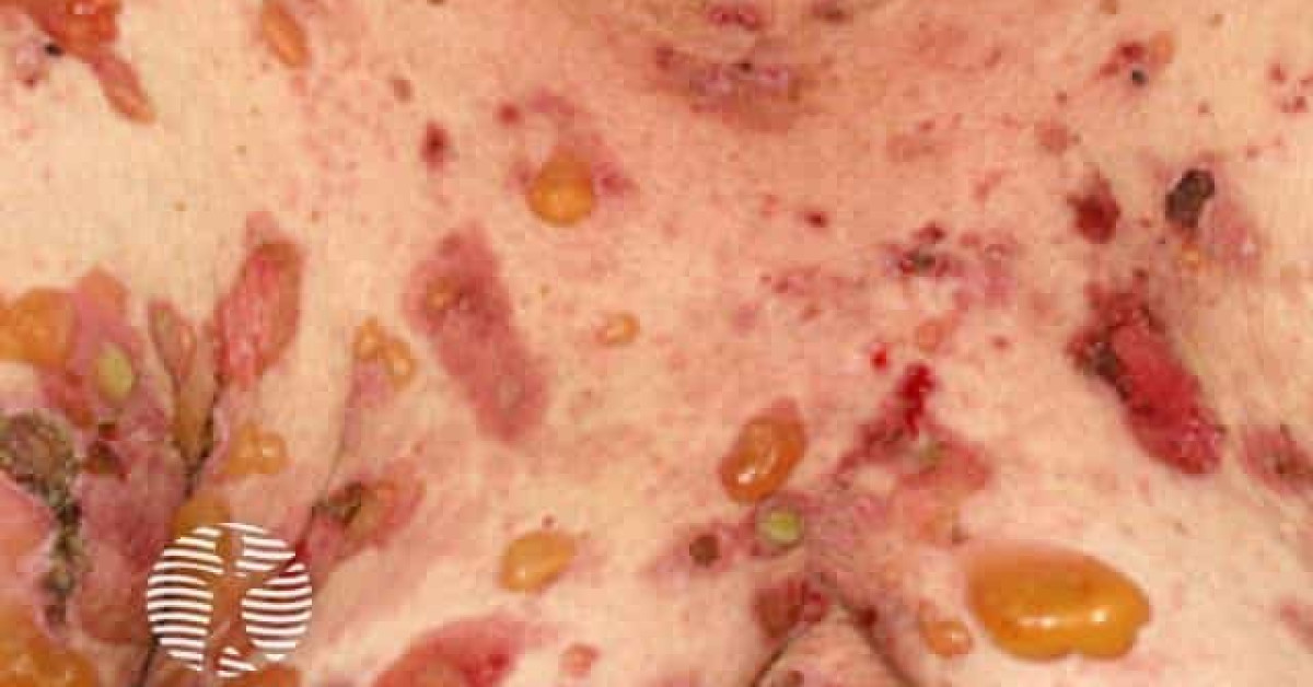
Bullous pemphigoid is the commonest autoimmune subepidermal blistering disease, affecting principally adults over 70. Autoantibodies to hemidesmosomal proteins BP180 (BPAG2) and BP230 produce tense bullae on erythematous or urticated bases. There is an established association with neurological disease and with antidiabetic gliptin therapy. Bullous pemphigoid is an increasingly recognised cutaneous immune-related adverse event of PD-1 / PD-L1 inhibitors, often emerging months after ICI initiation. EADV S2k guidance prioritises super-potent topical corticosteroids where feasible, with oral prednisolone, doxycycline-based strategies or steroid-sparing immunosuppression according to severity and comorbidity.
Epidemiology
- UK incidence ~4-5 per 100 000 / year and rising.
- Mean age >75; bilateral sex distribution.
- Associations: neurological disease (stroke, dementia, Parkinson), gliptins (vildagliptin > sitagliptin), spironolactone, furosemide; rare malignancy association (lung, GI, haematological).
- ICI-induced bullous pemphigoid: increasingly recognised G3-G4 cutaneous immune-related adverse event of PD-1 / PD-L1 inhibitors — onset typically 4-12 months after first dose; may persist after ICI discontinuation.
Clinical features
- Pre-bullous phase (weeks to months): pruritic urticarial / eczematous plaques.
- Bullous phase: tense bullae on erythematous or urticated bases; clear or haemorrhagic fluid; intact roof on rupture leaves erosion.
- Distribution: flexor forearms, axillae, groin, abdomen, lower legs; mucosal involvement less than mucous membrane pemphigoid (~10-30%).
- Nikolsky sign negative (vs pemphigus).
- Variants: localised, vesicular, nodular, vegetans, erythrodermic, dyshidrosiform.
Investigations
- Skin biopsy (lesional edge): subepidermal blister with eosinophil-rich infiltrate.
- Direct immunofluorescence (perilesional skin, transport medium): linear C3 ± IgG along basement membrane zone.
- Indirect immunofluorescence (serum) on salt-split skin: IgG binds the epidermal roof.
- ELISA anti-BP180 NC16A, anti-BP230 — confirm diagnosis and monitor disease activity.
- Investigate for triggers: drug history (gliptins!), neurological exam, malignancy screen if indicated.
Differential diagnosis
- Mucous membrane pemphigoid — mucosal predominance; scarring.
- Linear IgA disease — vancomycin-associated; younger.
- Pemphigus vulgaris — flaccid bullae, Nikolsky+, mucosal-first; DSG3 antibodies.
- Dermatitis herpetiformis — coeliac association; intensely pruritic vesicles, IgA at dermal papillae tips.
- Epidermolysis bullosa acquisita — extensor / trauma sites; IIF binds dermal floor.
- Bullous drug reaction, eczema with bullae, scabies (in elderly), bullous arthropod reaction.
Management (EADV 2022 / BAD 2012)
- First-line:
- Super-potent topical corticosteroid (clobetasol propionate 0.05% 30-40 g/day) over whole body or affected areas — French data (Joly et al, NEJM 2002) shows comparable efficacy and lower mortality than oral steroids.
- Oral doxycycline 200 mg OD is the BLISTER-trial alternative for mild-moderate disease.
- Second-line: oral prednisolone 0.3-0.5 mg/kg/day with steroid-sparing agent (methotrexate, azathioprine, mycophenolate mofetil).
- Refractory disease: rituximab, omalizumab, dupilumab; IVIG, plasma exchange in severe cases.
- ICI-induced BP: hold ICI for G3+; topical / oral steroids; doxycycline; rituximab or dupilumab in steroid-refractory cases. ICI rechallenge per cutaneous-irAE pathway.
- Stop suspected drug trigger; eczema-grade skin care (bland emollients, infection surveillance).
References
- Joly P et al. A comparison of oral and topical corticosteroids in patients with bullous pemphigoid. N Engl J Med. 2002;346:321-327.
- Williams HC et al. Doxycycline versus prednisolone as an initial treatment strategy for bullous pemphigoid (BLISTER). Lancet. 2017;389:1630-1638.
- Borradori L et al. Updated S2k guidelines for the management of bullous pemphigoid initiated by the European Academy of Dermatology and Venereology (EADV). J Eur Acad Dermatol Venereol. 2022;36(10):1689-1704.
- Venning VA et al. British Association of Dermatologists' guidelines for the management of bullous pemphigoid 2012. Br J Dermatol. 2012;167(6):1200-1214.
- Sibaud V et al. Bullous pemphigoid associated with immune checkpoint inhibitors: clinical features and management. Br J Dermatol. 2021;185:1003-1011.
- Schulze F et al. Bullous pemphigoid in dipeptidyl peptidase-4 inhibitor users: a population-based cohort study. JAMA Dermatol. 2018;154:1152-1158.
Spot a correction?
If any clinical statement, citation or link on this page needs updating, please email admin@skinoncology.net with the page name, the proposed correction and the supporting source.