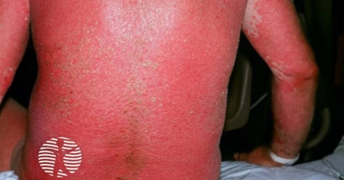
Erythroderma
Exfoliative dermatitis · generalised exfoliative erythroderma
Erythroderma denotes generalised erythema affecting ≥90% body-surface area, often with scaling, that develops over weeks to months. It is a clinical syndrome, not a diagnosis, with major aetiologies including drug reactions, pre-existing dermatoses (psoriasis, atopic eczema, pityriasis rubra pilaris), cutaneous T-cell lymphoma (Sézary, mycosis fungoides), other internal malignancies and idiopathic disease. Up to 20% are paraneoplastic. Management is a dermatologic emergency requiring inpatient supportive care alongside an aetiological workup.
Aetiology
- Pre-existing dermatoses (~50%): psoriasis, atopic eczema, pityriasis rubra pilaris, contact dermatitis, drug eruption flare.
- Drug reaction (~10-30%): anticonvulsants (lamotrigine, carbamazepine), allopurinol, sulfonamides, NSAIDs, antibiotics, ICIs.
- Cutaneous T-cell lymphoma (~15-20%): Sézary syndrome, erythrodermic mycosis fungoides.
- Other malignancy (~5-10%): solid-organ (lung, GI, prostate, breast), other lymphomas / leukaemias.
- Idiopathic (~10-20%) — red-man syndrome; older men; consider occult CTCL.
- Infection: staphylococcal scalded-skin (paediatric), exanthem.
Clinical features
- Erythema and scaling involving ≥90% BSA.
- Pruritus; lichenification in chronic phase.
- Lymphadenopathy (reactive / dermatopathic; rule out lymphomatous spread).
- Diffuse non-scarring alopecia, palmoplantar keratoderma, ectropion, nail dystrophy.
- Systemic effects: hypothermia, fluid loss, hypoalbuminaemia, high-output cardiac failure, oedema, electrolyte imbalance, sepsis.
Workup
- Skin biopsy — 2-3 sites; may need repeated biopsies as histology often non-specific.
- Bloods: FBC with film, peripheral blood flow cytometry (Sézary cells / B-cell clone), LDH, U&E, LFT, albumin, IgE, HIV / HepBC, ferritin.
- T-cell receptor gene rearrangement in skin and blood if CTCL suspected.
- Imaging — CT chest / abdomen / pelvis if lymphadenopathy, B-symptoms or no obvious aetiology.
- Patch testing if contact / drug aetiology suspected (after stabilisation).
- Repeated drug-history review and discontinuation of all non-essential medications.
Management
- Hospital admission for any patient with haemodynamic, thermoregulatory or electrolyte instability; bed-rest in a warm environment with bland emollients.
- IV fluids, electrolyte correction, calorie / protein replacement, antimicrobials for secondary infection.
- Wet dressings, mid-potency topical steroids; avoid potent topical steroids over erythrodermic skin (systemic absorption).
- Aetiology-directed: stop the offending drug; treat underlying psoriasis (acitretin, methotrexate, ciclosporin, biologics); CTCL (ECP, mogamulizumab, RT, brentuximab); treat occult malignancy.
- Critical-care or burns-unit support for severe cases.
Practical points
- Always re-biopsy if no diagnosis after 6-12 months — CTCL often unmasks itself with time.
- Peripheral-blood flow cytometry for Sézary cells (CD4+ CD7− / CD26−) at presentation and follow-up.
- Hold immunotherapy in suspected ICI-related erythroderma; corticosteroids; multidisciplinary discussion before re-challenge.
- Counsel patients about long-term risk of recurrence and need for skin examination.
References
- Cuellar-Barboza A et al. Erythroderma: a comprehensive review. Am J Clin Dermatol. 2024;25:367-379.
- Mistry N et al. A review of erythroderma. Br J Hosp Med. 2015;76:C49-C52.
- Sigurdsson V et al. Erythroderma. A clinical and follow-up study of 102 patients. J Am Acad Dermatol. 1996;35:53-57.
- British Association of Dermatologists. Erythroderma — patient information leaflet (clinical guidelines digest). London: BAD; 2022.
Spot a correction?
If any clinical statement, citation or link on this page needs updating, please email admin@skinoncology.net with the page name, the proposed correction and the supporting source.