

Atrophie blanche / livedoid vasculopathy
Livedoid vasculopathy; LV; LVL; PURPLE syndrome; atrophie blanche en plaque; segmental hyalinising vasculitis
Atrophie blanche — properly named livedoid vasculopathy — is a chronic small-vessel thrombotic vasculopathy of the lower limbs presenting as recurrent painful ulcers that heal with characteristic porcelain-white stellate atrophic scars ("atrophie blanche"). Pathogenesis involves dermal-vessel thrombosis with secondary segmental hyalinisation. Hypercoagulable disorders (antiphospholipid syndrome, factor V Leiden, prothrombin G20210A, MTHFR, protein C / S / antithrombin deficiency) underlie a substantial proportion of cases. Clinically important as a chronic-ulcer differential in skin oncology — patients undergo repeated biopsies as suspected cSCC or vasculitis before recognition. Treatment is antiplatelet + anticoagulant therapy with adjunctive measures.
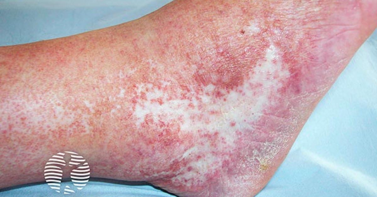
Clinical features
- Painful, recurrent ulcers of the lower legs and ankles, particularly the malleoli.
- Preceding livedo reticularis / racemosa pattern of mottled discoloration.
- Heals with characteristic porcelain-white stellate atrophic scars ringed by telangiectasia and hyperpigmentation — "atrophie blanche".
- Chronic relapsing-remitting course over years; seasonal worsening (winter) common.
- Female predominance; peak age 30–50.
- Severity ranges from minor recurrent ulceration to disabling chronic disease.
Pathogenesis
- Primary thrombotic vasculopathy of small dermal vessels (50–200 µm) with secondary segmental hyalinisation of vessel walls.
- Not primarily inflammatory — distinguishes from true vasculitis.
- Hypercoagulable predisposition in up to ~50% (reported prevalence varies widely across series):
- Antiphospholipid syndrome.
- Factor V Leiden, prothrombin G20210A.
- MTHFR mutations / hyperhomocysteinaemia.
- Protein C, S, antithrombin deficiency.
- Plasminogen-activator inhibitor-1 (PAI-1) elevation.
- Cryoglobulinaemia.
- Idiopathic in the remainder.
Diagnosis
- Clinical pattern — recurrent lower-limb ulceration with porcelain-white scarring — is highly suggestive.
- Biopsy of ulcer edge:
- Hyalinised, thickened small-vessel walls; intraluminal fibrin thrombi.
- Minimal vasculitic inflammation in true LV (distinguishes from true vasculitis).
- Direct immunofluorescence — limited specific findings.
- Hypercoagulability screen — lupus anticoagulant, anticardiolipin antibodies, β2-GP1, factor V Leiden, prothrombin G20210A, MTHFR, homocysteine, protein C / S / antithrombin, cryoglobulins.
- Doppler ultrasound — exclude chronic venous insufficiency / arterial disease.
- Multidisciplinary input — dermatology, haematology, vascular medicine.
Management
- Antithrombotic therapy is the cornerstone:
- Aspirin 75–300 mg daily.
- Anticoagulation — rivaroxaban 10–20 mg daily is increasingly used (open-label RILIVAS data; robust RCT evidence is limited); alternatives include warfarin, LMWH, dabigatran.
- Combination antiplatelet + anticoagulant for severe disease.
- Adjunctive therapy:
- Pentoxifylline 400 mg TDS — modest evidence for ulcer healing.
- Hyperbaric oxygen therapy in refractory cases.
- IVIg for cases unresponsive to anticoagulation.
- PUVA, topical tacrolimus — anecdotal benefit.
- Hypercoagulable treatment — anticoagulation in APS, B-vitamin / folate supplementation in hyperhomocysteinaemia.
- Wound care — compression hosiery, non-adherent dressings, infection management.
- Pain control — substantial; nociceptive + neuropathic components.
- Sun protection of fragile scarring skin.
Prognosis
- Chronic relapsing course over years; rarely entirely resolved.
- Excellent overall prognosis but significant chronic morbidity from pain and recurrent ulceration.
- Effective antithrombotic therapy substantially reduces recurrence frequency in 60–80% of patients.
- Underlying hypercoagulability often requires lifelong anticoagulation.
References
- Kerk N, Goerge T. Livedoid vasculopathy — current aspects of diagnosis and treatment. J Dtsch Dermatol Ges; 2013.
- Weishaupt C et al. Rivaroxaban for the treatment of livedoid vasculopathy (RILIVAS). Lancet Haematol; 2016.
Spot a correction?
If any clinical statement, citation or link on this page needs updating, please email admin@skinoncology.net with the page name, the proposed correction and the supporting source.