Necrobiosis lipoidica
NL; necrobiosis lipoidica diabeticorum (older — only ~50% are diabetic); "shin spot of diabetes" (informal)
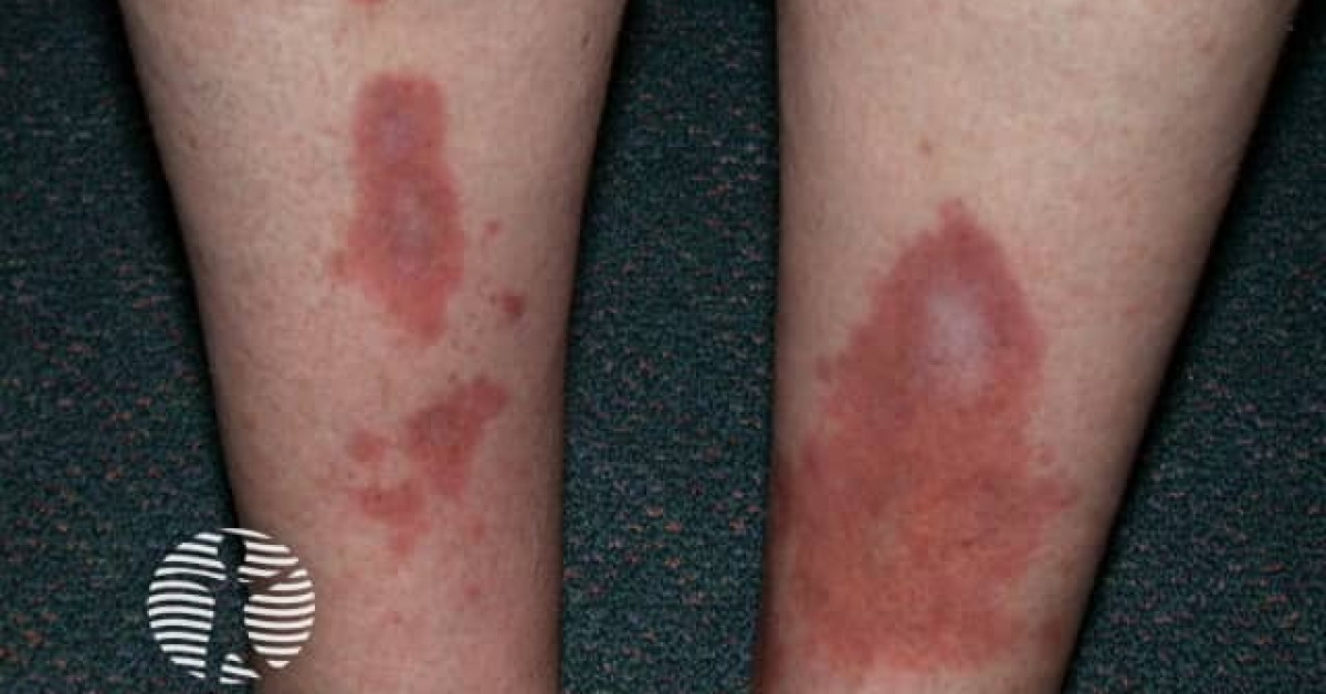
Necrobiosis lipoidica is a chronic granulomatous dermatosis classically presenting as a slowly enlarging atrophic, telangiectatic, yellow-brown plaque on the pretibial region of a young to middle-aged adult. Approximately 50% of patients have diabetes mellitus and a further 10% develop diabetes during follow-up — making screening for impaired glucose tolerance mandatory. The disease has skin oncology relevance because chronic ulceration of long-standing necrobiosis lipoidica plaques is a recognised substrate for Marjolin-spectrum squamous cell carcinoma, though this is rare and reported largely at case-report level in long-standing ulcerated lesions. The combination of chronic inflammation, scarring, ulceration and impaired wound healing in diabetic patients further amplifies the risk. Surveillance for change in long-standing lesions, low threshold for biopsy of any non-healing ulcer or new induration, and optimised inflammatory control are the key preventive interventions.
Clinical features
- Slowly enlarging, well-demarcated yellow-brown to red-brown plaques with an atrophic, shiny, "cellophane"-thin centre and a raised inflammatory edge.
- Prominent telangiectasias visible through the atrophic surface.
- Distribution — pretibial (~85%), occasionally on dorsal feet, arms, scalp, face, abdomen.
- Often bilateral and symmetric.
- Onset typically 30–50 years; F:M ~3:1.
- ~50% have diabetes mellitus (type 1 > type 2) at diagnosis; a further 10% develop diabetes during follow-up.
- Asymptomatic or mildly itchy / sore; significant pain only when ulcerated.
- Ulceration in ~30% of long-standing lesions — frequently triggered by minor trauma; chronic, painful, slow to heal.
- Differential — granuloma annulare (especially the disseminated variant), sarcoidosis, lichen sclerosus, lipodermatosclerosis, morphea, basal cell carcinoma (within long-standing ulcerated NL).
Histology
- Necrobiotic ("rotten") collagen in horizontal layers throughout the dermis, alternating with palisading granulomas of histiocytes and lymphocytes ("layered" or "lasagne" pattern, contrasting with the focal palisade of granuloma annulare).
- Microvascular changes — endothelial thickening, perivascular inflammation, sclerosis.
- Plasma cells and lipid deposition (lipid-laden histiocytes / cholesterol clefts) — yellow-brown clinical colour.
- Loss of elastic fibres in the centre of the plaque.
- If ulcerated and chronic — biopsy should sample the ulcer base / edge to exclude SCC arising in NL.
SCC arising in necrobiosis lipoidica
- SCC in long-standing necrobiosis lipoidica plaques is rare and reported largely at case-report level (no robust denominator); risk amplified by chronic ulceration, immunosuppression and diabetic microvascular impairment.
- Behaves as a Marjolin-spectrum SCC — see monograph.
- Latency typically 20–30 years from NL onset to SCC diagnosis.
- Warning signs:
- New non-healing ulcer or rapidly enlarging exophytic mass within an established NL plaque.
- Induration disproportionate to the surrounding plaque.
- Pain disproportionate to the patient's chronic baseline.
- Bleeding.
- Multiple deep biopsies of any change; manage as Marjolin SCC.
- Less commonly reported — basal cell carcinoma arising in NL.
Management
- Diabetes management — confirm or exclude diabetes; optimise glycaemic control (does not directly resolve NL but reduces complications).
- First-line skin therapy:
- Potent / ultrapotent topical corticosteroid for the active inflammatory edge (avoid prolonged centrally — accelerates atrophy).
- Intralesional triamcinolone for refractory peripheral inflammation.
- Topical calcineurin inhibitor (tacrolimus, pimecrolimus).
- Refractory disease:
- Pentoxifylline.
- PUVA / topical PUVA / UVA1 phototherapy.
- Hydroxychloroquine.
- JAK inhibitors (case reports — tofacitinib, baricitinib).
- TNF inhibitors (infliximab, etanercept, adalimumab) for refractory ulcerated disease.
- Surgical excision and grafting for ulcerated, refractory, scarring lesions in selected cases (high risk of recurrence in graft margins).
- Ulcer management — wound care, compression, infection management, pain control, dietitian.
- Cancer surveillance — annual review with photographic documentation; biopsy any change.
- Multidisciplinary involvement — diabetes specialist, vascular medicine, plastic surgery for ulceration / SCC.
References
- Reid SD et al. Update on necrobiosis lipoidica — review. J Am Acad Dermatol; 2013.
- Patel GK, Harding KG. Squamous cell carcinoma in long-standing necrobiosis lipoidica. J Wound Care; 2013.
Spot a correction?
If any clinical statement, citation or link on this page needs updating, please email admin@skinoncology.net with the page name, the proposed correction and the supporting source.