

Milia
Milium (singular) / milia (plural); "milk spots"; congenital milia (infantile); secondary / "post-traumatic" milia
Milia are small (1–3 mm), firm, dome-shaped, white-yellow superficial epidermal keratin-filled cysts. Two clinical contexts are recognised: primary milia (idiopathic, including infantile milia, the multiple eruptive milia of adolescence, and the milia en plaque variant) — generally appearing on the face (cheeks, eyelids, periorbital, forehead) of healthy individuals; and secondary milia — arising at sites of previous blistering disease (epidermolysis bullosa, porphyria cutanea tarda, bullous pemphigoid, second-degree burns), trauma (dermabrasion, chemical peel, laser resurfacing) or chronic topical corticosteroid use. Milia are entirely benign with no malignant potential. The skin-oncology relevance is twofold: (1) clinical confusion with sebaceous hyperplasia, syringoma, BCC, trichoepithelioma or other small facial tumours; and (2) the recognition that multiple milia in a young patient with other cutaneous features can be an early marker of Bazex-Dupré-Christol (with hypotrichosis, follicular atrophoderma, multiple BCCs), Rombo syndrome (with vermiculate atrophoderma), Brooke-Spiegler syndrome or rare congenital genodermatoses (Gardner syndrome, basaloid follicular hamartoma syndrome).
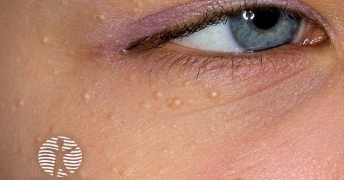
Clinical features
- Multiple small (1–3 mm), firm, dome-shaped, white to pearly-yellow superficial papules.
- Distribution — face (cheeks, eyelids, periorbital, forehead, nose); occasionally chest, back, genitalia.
- Asymptomatic; cosmetic concern is the principal presenting issue.
- Primary milia:
- Congenital / infantile milia — present at birth in 40–50% of full-term newborns; spontaneously resolve over weeks; nasal, perinasal, palatal (Epstein pearls).
- Multiple eruptive milia — multiple new milia in adolescence / young adult.
- Milia en plaque — clusters of milia within an erythematous plaque, classically post-auricular.
- Secondary milia:
- Following bullous disease — epidermolysis bullosa, porphyria cutanea tarda, bullous pemphigoid, epidermolysis bullosa acquisita (EBA), second-degree burns.
- Following trauma — dermabrasion, chemical peel, laser resurfacing, cryotherapy, contact dermatitis healing.
- Following chronic topical corticosteroid use.
- Following radiotherapy.
Histology
- Small (~1–2 mm) round superficial epidermal cyst with a thin stratified squamous epithelial wall (with granular layer) and a central keratin core.
- Located in the superficial dermis (in contrast to epidermoid cysts which are deeper).
- Usually arises from the infundibulum of the vellus hair follicle (primary milia) or from eccrine ducts / occluded follicles in damaged skin (secondary milia).
- No atypia.
Syndromic associations
- Multiple milia in a young patient with other cutaneous features — consider:
- Bazex-Dupré-Christol syndrome — milia + congenital hypotrichosis + follicular atrophoderma of the dorsal hands + multiple early BCCs — see monograph.
- Rombo syndrome — milia + vermiculate atrophoderma of the cheeks + hypotrichosis + peripheral cyanosis + multiple BCCs — see monograph.
- Brooke-Spiegler syndrome — milia in the context of multiple cylindromas, spiradenomas and trichoepitheliomas — see monograph.
- Gardner syndrome — multiple epidermoid cysts (sometimes with milia) in unusual sites + osteomas + colorectal polyposis (FAP) — see epidermoid cyst page.
- Basaloid follicular hamartoma syndrome — multiple basaloid follicular hamartomas + milia + alopecia.
- Atrichia with papular lesions — milia-like papules + alopecia.
- Congenital epidermolysis bullosa — bullous skin + multiple secondary milia at healed blister sites.
Management
- Reassurance — typical milia are benign and often spontaneously resolve (especially infantile).
- Removal options (cosmetic):
- Incision and expression with a hypodermic needle — first-line for individual milia; quick, simple, leaves no scar.
- Curettage with electrodesiccation.
- Cryotherapy.
- Topical retinoids (tretinoin) — for multiple eruptive milia; reduces formation rate.
- Oral retinoids (isotretinoin) — refractory multiple milia.
- CO₂ laser ablation — milia en plaque.
- Histology unnecessary for typical milia; biopsy if any diagnostic doubt or syndromic context.
- Identify and treat any underlying bullous disease, drug, trauma or syndromic context.
References
- Berk DR, Bayliss SJ. Milia — a review and classification. J Am Acad Dermatol; 2008.
- Tsuruta D et al. Milia en plaque — review. J Am Acad Dermatol; 2008.
Spot a correction?
If any clinical statement, citation or link on this page needs updating, please email admin@skinoncology.net with the page name, the proposed correction and the supporting source.