Lymphoedema (general overview)
Primary lymphoedema · secondary lymphoedema · lymphatic insufficiency
Lymphoedema is a chronic limb / regional swelling caused by impaired lymphatic drainage. It is classified as primary (congenital / genetic — Milroy disease, Meige disease, lymphoedema-distichiasis) or secondary (most commonly after lymphadenectomy, radiotherapy, infection — filariasis — or trauma). In UK skin-oncology practice it predominantly follows melanoma / breast / vulval-cancer lymphadenectomy. Diagnosis is clinical, supplemented by ISL staging, lymphoscintigraphy and ICG lymphography. Modern management is multidisciplinary — complex decongestive therapy (CDT), compression, and supermicrosurgical reconstruction (LVA, VLNT).
Classification
- Primary lymphoedema:
- Congenital (Milroy disease, FLT4/VEGFR3 mutation) — present at birth.
- Lymphoedema praecox (Meige) — onset puberty - 35 years.
- Lymphoedema tarda — onset >35 years.
- Syndromic: lymphoedema-distichiasis (FOXC2), Turner, Noonan, Hennekam.
- Secondary lymphoedema:
- Post-oncology: lymphadenectomy (breast / melanoma / vulval / penile / gynae), radiotherapy.
- Filarial: Wuchereria bancrofti in endemic countries (commonest globally).
- Trauma, infection (recurrent cellulitis).
- Tumour-related lymphatic obstruction (cutaneous metastases, advanced cancer).
- Chronic venous insufficiency overlap (phlebolymphoedema).
- Obesity-related lymphoedema.
ISL staging (2020)
- Stage 0 (latent): subclinical; impaired transport demonstrable on lymphoscintigraphy.
- Stage I: pitting oedema reversed by elevation.
- Stage II: progressive fibrosis; reduced pitting; not fully reversible.
- Stage III: lymphostatic elephantiasis with trophic skin changes, hyperkeratosis, papillomatosis.
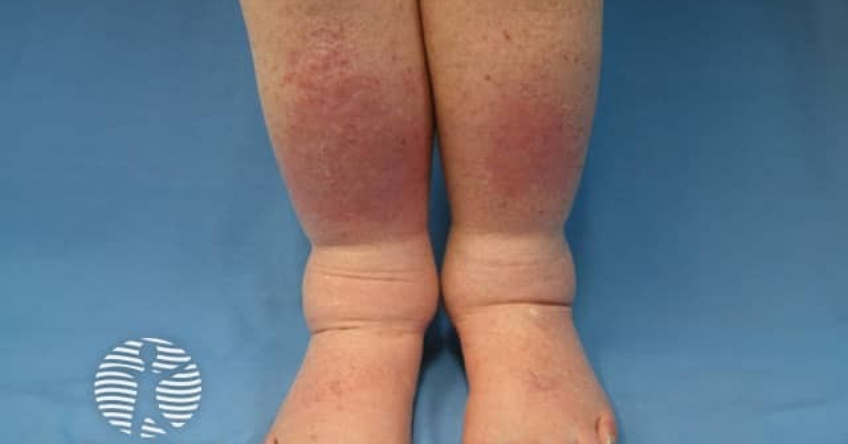
Clinical features
- Painless heavy swelling; usually unilateral.
- Stemmer sign positive (cannot pinch second toe / finger fold).
- Skin changes: peau d'orange, hyperkeratosis, lymphorrhoea, papillomatosis.
- Recurrent cellulitis episodes.
- Functional limitation, body-image issues.
- Complications:
- Recurrent cellulitis (each episode worsens lymphoedema).
- Cutaneous angiosarcoma (Stewart-Treves syndrome) — rare but important; persistent / chronic lymphoedema risk; bluish nodules / patches; refer urgently for biopsy.
- Functional impairment, depression.
Workup
- Limb circumference measurement; perometer / water displacement.
- Bioimpedance spectroscopy (L-Dex) — early latent disease.
- Lymphoscintigraphy — gold standard imaging.
- ICG lymphography — superficial lymphatics; preoperative LVA planning.
- MR lymphangiography — complex cases.
- USS Doppler — exclude DVT / venous component.
- Bloods if filaria endemic country: blood film, microfilarial PCR.
- Genetic testing for primary lymphoedema (FLT4, FOXC2 — R124 / R142 gene panels).
Management
- Complex decongestive therapy (CDT) — first-line:
- Manual lymphatic drainage (MLD).
- Multilayer short-stretch compression bandaging.
- Compression garments (class 1-3) fitted by specialist.
- Skin care: emollients, infection prevention.
- Decongestive exercise.
- Pneumatic compression devices — adjunctive.
- Pharmacology: limited evidence; coumarin / selenium tried historically; not routinely recommended.
- Surgical (specialist centres):
- Lymphaticovenular anastomosis (LVA) — early-stage; supermicrosurgical anastomosis of lymphatic to venule.
- Vascularised lymph-node transfer (VLNT) — flap-based donor lymph-node transfer to affected basin.
- Suction-assisted lipectomy / debulking — late-stage fibrofatty disease.
- Antibiotic prophylaxis: phenoxymethylpenicillin 250 mg BD long-term for ≥2 cellulitis episodes / 12 months (PATCH-II).
- Multidisciplinary: lymphoedema clinic, physiotherapy, plastic surgery, dermatology, vascular, psychology, dietetics.
References
- International Society of Lymphology. Diagnosis and treatment of peripheral lymphedema: 2020 consensus document. Lymphology. 2020;53:3-19.
- British Lymphology Society. Standards of practice for the management of lymphoedema. Sevenoaks: BLS; 2023.
- Schaverien MV et al. Lymphedema: state-of-the-art review. Plast Reconstr Surg. 2017;140:1003e-1017e.
- Lymphoedema Support Network (LSN). UK patient resource. London: LSN; 2024.
- NICE NG101. Early and locally advanced breast cancer: diagnosis and management. London: NICE; 2018 (last updated 14 April 2025), recommendations 1.14.8-1.14.11.
- NICE HTG622. Liposuction for chronic lymphoedema. London: NICE; 2022.
- NICE HTG717. Lymphovenous anastomosis during axillary or inguinal node dissection for preventing secondary lymphoedema. London: NICE; 2024.
Spot a correction?
If any clinical statement, citation or link on this page needs updating, please email admin@skinoncology.net with the page name, the proposed correction and the supporting source.