

GenodermatosisX-linked dominantOMIM 308300
Incontinentia pigmenti
IP · Bloch-Sulzberger syndrome · IKBKG / NEMO disease
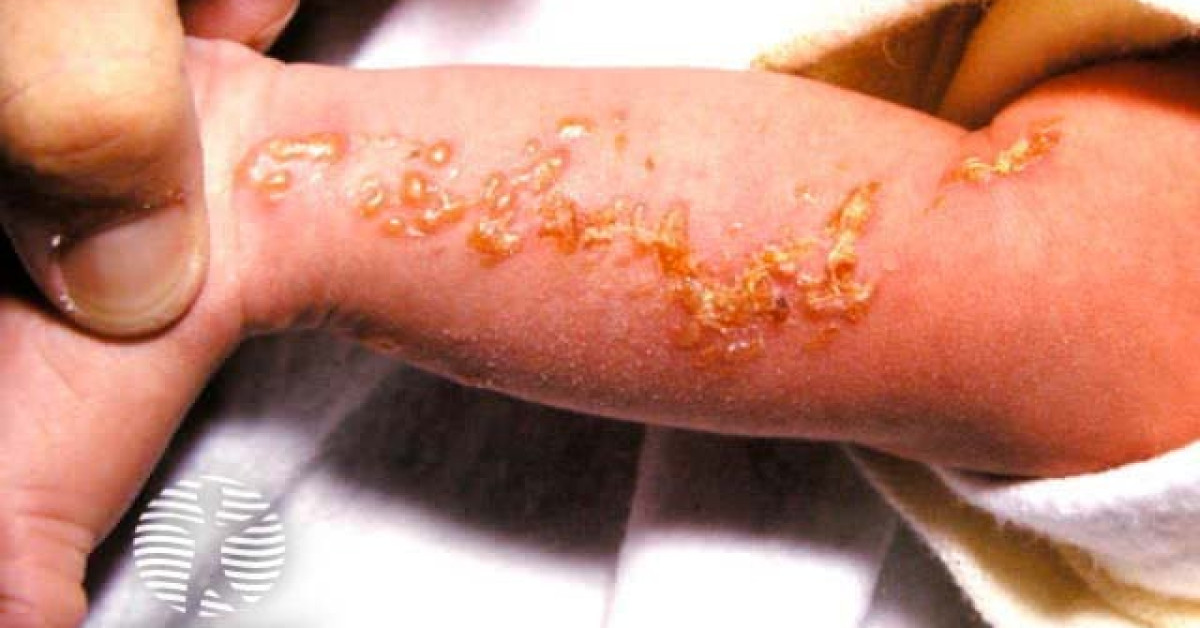
Incontinentia pigmenti is an X-linked dominant ectodermal genodermatosis caused by mutations in IKBKG (NEMO), affecting NF-κB pathway signalling. The classical four-stage cutaneous evolution — vesicular, verrucous, hyperpigmented (Blaschko-linear), atrophic / hypopigmented — typically begins in the neonatal period. Female predominance reflects in-utero lethality in most affected males. Associated CNS, ocular, dental and skeletal features require multidisciplinary surveillance.
CurrentLast reviewed 16 May 2026
Genetics
- X-linked dominant; IKBKG (NEMO) on Xq28.
- Affected females mosaic; affected males rare and survive only with Klinefelter (XXY) or mosaicism.
- Female : male ratio ~20:1.
- Most cases are de novo; family history in some.
- NF-κB signalling defect → apoptosis susceptibility, ectodermal dysplasia features.
Four cutaneous stages
- Stage 1 — Vesicular (birth to 4 months):
- Linear erythematous vesicles / bullae along Blaschko lines.
- Peripheral eosinophilia common.
- Resembles bullous impetigo / HSV.
- Stage 2 — Verrucous (2-6 months):
- Linear hyperkeratotic verrucous plaques.
- Resolves over months.
- Stage 3 — Hyperpigmented (most characteristic) (3-6 months to puberty):
- Brown swirled / whorled pigmentation along Blaschko lines.
- Trunk, limbs predominant.
- Stage 4 — Atrophic / hypopigmented (adolescence onwards):
- Pale, atrophic, sometimes hairless streaks along Blaschko lines.
- Often subtle in adults; lifelong.
Systemic features
- CNS (~30%): seizures, developmental delay, spasticity, hemiparesis; vasculopathy.
- Ocular (~35%): retinal vascular abnormalities, retinal detachment, blindness; cataract, optic atrophy.
- Dental: hypodontia, peg / conical teeth, microdontia, delayed eruption (~80%).
- Hair: alopecia, vertex pseudopelade-like patches.
- Nails: dystrophy, ridging.
- Skeletal: rare.
- Increased keratinocyte malignancy: not classically described but case reports of cSCC in Stage 4 plaques in older patients.
Investigations
- Clinical diagnosis at vesicular / verrucous stage with characteristic distribution.
- Skin biopsy: eosinophilic spongiosis (Stage 1); dyskeratosis; pigment incontinence (melanophages) in Stage 3.
- FBC: eosinophilia in early stages.
- Genetic confirmation: IKBKG sequencing (R203 panel).
- Ophthalmology review: urgent paediatric ophthalmology; serial retinal exams; consider EUA / RetCam.
- Neurology / brain MRI for any neurological symptom.
- Dental: paediatric dental review.
- Karyotype if affected male (consider Klinefelter).
Management
- Skin care:
- Stage 1: gentle bland emollient; avoid secondary infection (DDx HSV — viral swabs / aciclovir if clinical concern).
- Stage 2: emollient; no specific treatment.
- Stage 3: cosmetic; pigment fades over years.
- Stage 4: cosmetic; sun protection.
- Ophthalmology: laser photocoagulation for proliferative retinopathy; vitreoretinal surgery.
- Neurology: anti-epileptic management; developmental support.
- Dental: prosthodontic management.
- Genetic counselling: 50% transmission risk; PIGD / chorionic villus sampling option for affected mothers.
- Long-term multidisciplinary follow-up: dermatology, ophthalmology, neurology, dental, genetics.
References
- Bloch B. Eigentümliche, bisher nicht beschriebene Pigmentaffektion (incontinentia pigmenti). Schweiz Med Wochenschr. 1926;56:404-407.
- Smahi A et al. Genomic rearrangement in NEMO impairs NF-kappaB activation and is a cause of incontinentia pigmenti. Nature. 2000;405:466-472.
- Minić S et al. Systematic review of incontinentia pigmenti: 920 cases. Clin Genet. 2014;85:536-542.
- Greene-Roethke C. Incontinentia pigmenti: a summary review. J Pediatr Health Care. 2017;31:e45-e52.
Spot a correction?
If any clinical statement, citation or link on this page needs updating, please email admin@skinoncology.net with the page name, the proposed correction and the supporting source.