Granular cell tumour
Abrikossoff tumour; granular cell myoblastoma (older, incorrect — they are Schwann-cell, not muscle-cell, in origin)
Granular cell tumour is an uncommon soft-tissue tumour of Schwann-cell origin (S100-positive) characterised by sheets of polygonal cells with abundant granular eosinophilic cytoplasm. The tongue is the commonest single site (~30%), followed by skin and subcutis (~30%) — particularly head, neck, breast and proximal upper limb — with a smaller proportion in the gastrointestinal tract, breast parenchyma and respiratory tree. The vast majority (~98%) are benign, behaving as a slow-growing, indolent dermal/subcutaneous nodule. The malignant variant is rare (<2%) but aggressive, with high rates of regional and distant metastasis. Skin biopsy frequently shows florid pseudoepitheliomatous hyperplasia of the overlying epidermis that can be misdiagnosed as squamous cell carcinoma — a critical pitfall that requires the histopathologist to recognise the underlying granular cells.
Clinical features
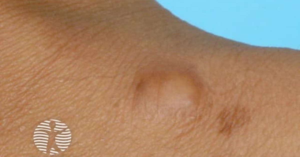
- Solitary firm, skin-coloured to pink/yellow dermal/subcutaneous nodule, usually 1–3 cm.
- Site distribution — tongue ~30%, skin/subcutis ~30% (head/neck, breast, proximal upper limb), GI tract, breast, lung.
- Median age 30–60; F>M (~2:1); commoner in patients of African descent.
- Multiple lesions in 5–25%; can be associated with rare syndromes (Noonan, LEOPARD, neurofibromatosis 1).
- Asymptomatic; may itch or feel tethered to deeper structures.
Histology & pitfalls
- Sheets and nests of large polygonal cells with abundant eosinophilic granular cytoplasm and small bland nuclei.
- PAS-positive, diastase-resistant cytoplasmic granules (lysosomes).
- Strongly S100, SOX10, CD68, NKI/C3 positive — confirming Schwann-cell origin.
- Frequently shows florid pseudoepitheliomatous hyperplasia of the overlying epidermis — a common cause of misdiagnosis as well-differentiated SCC on superficial biopsy. Always include a deep biopsy if granular cell tumour is suspected; the granular cell component is in the dermis.
- Fanburg-Smith criteria for malignancy (≥3 of 6 features = malignant; 1–2 = atypical):
- Necrosis
- Spindling
- Vesicular nuclei with prominent nucleoli
- High N:C ratio
- Mitotic count >2/10 HPF
- Pleomorphism
Management
- Benign granular cell tumour: complete surgical excision with narrow (3–5 mm) margins; recurrence ~5% with incomplete excision.
- Atypical or malignant granular cell tumour: wide local excision with 2 cm margins; sentinel lymph node biopsy considered; referral to sarcoma MDT.
- Imaging staging (CT chest/abdomen/pelvis) for atypical/malignant disease.
- Adjuvant radiotherapy for incomplete margins or high-risk disease.
- Systemic therapy for metastatic disease — limited evidence; multikinase inhibitors (pazopanib), mTOR inhibitors and case reports of immune checkpoint activity.
Prognosis
Benign GCT — excellent; cure with complete excision. Malignant GCT — 5-year overall survival 30–60%; high local recurrence and distant metastasis (lung, liver, bone, lymph nodes). Long-term surveillance for any atypical or malignant lesion.
References
- Fanburg-Smith JC et al. Malignant granular cell tumor of soft tissue: diagnostic criteria and clinicopathologic correlation. Am J Surg Pathol; 1998.
- Vered M et al. Granular cell tumor of the oral cavity: a clinicopathological study. J Oral Pathol Med; 2009.
Spot a correction?
If any clinical statement, citation or link on this page needs updating, please email admin@skinoncology.net with the page name, the proposed correction and the supporting source.