

Discoid lupus erythematosus
Chronic cutaneous lupus erythematosus; CCLE; chronic discoid LE; cutaneous lupus
Discoid lupus erythematosus is the commonest form of chronic cutaneous lupus erythematosus — a scarring inflammatory dermatosis presenting as well-demarcated erythematous plaques with follicular plugging, scale, atrophy and dyspigmentation, classically on photo-exposed skin (face, scalp, ears, dorsal hands, V of the neck). Approximately 5–10% of DLE patients develop systemic lupus erythematosus (SLE). DLE is an important skin-oncology entity because long-standing DLE plaques — particularly on the scalp and lower lip — develop aggressive cutaneous SCC in approximately 2–4% of patients (Marjolin-like). Diagnostic biopsy and ANA screening guide management. UK practice follows BAD guidance, with hydroxychloroquine as the cornerstone systemic therapy.
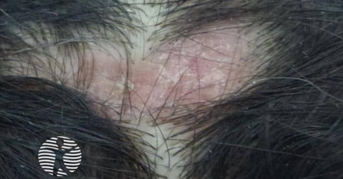
Clinical features
- Well-demarcated erythematous plaque with overlying adherent scale that on removal shows underside "tin-tack" follicular plugging.
- Follicular plugging — characteristic on dermoscopy and histology.
- Lesions evolve to central atrophy, dyspigmentation (hyper- and hypo-) and scarring.
- Sites — face (cheeks, ears, nose), scalp (scarring alopecia), V of the neck, dorsal hands.
- Hypertrophic / verrucous DLE — thickened keratotic variant; can mimic cSCC or KA.
- Mucosal DLE — oral lichenoid plaques; lip vermilion (cheilitis) with high SCC risk.
- Tumid LE — non-scarring lupus variant; not strictly DLE.
Progression to systemic LE
- 5–10% of patients with apparently isolated DLE develop SLE over time.
- Risk factors — generalised DLE (above and below the neck), positive ANA at presentation, female sex.
- Annual systemic screening — ANA, dsDNA, ENA, complement, urinalysis; symptom enquiry.
- Refer to rheumatology if SLE features emerge or screening abnormal.
cSCC risk in chronic DLE (DLE-associated SCC)
- Long-standing scarring DLE develops cutaneous SCC in approximately 2–4% of patients.
- Highest risk sites — chronically inflamed scarring scalp plaques and chronic discoid cheilitis of the lower lip.
- SCC arising in DLE behaves aggressively — higher recurrence and metastatic rates than usual cSCC.
- Photoprotection, smoking cessation and tight DLE disease control reduce risk.
- Biopsy any new or non-healing area within a stable DLE plaque.
Diagnosis
- Skin biopsy (lesional, edge):
- Vacuolar interface dermatitis with apoptotic keratinocytes.
- Perifollicular and perivascular lymphocytic infiltrate; follicular plugging; mucin in the dermis.
- Atrophic epidermis with basement-membrane thickening (PAS+).
- Direct immunofluorescence — granular IgG / IgM / C3 at the dermoepidermal junction (lupus band).
- Bloods — FBC, ANA, ENA panel, dsDNA, complement C3 / C4, RF, urinalysis.
- Photoprovocation testing in selected cases.
Management
- Photoprotection — SPF 50+ broad-spectrum daily; UV-protective clothing; sun avoidance during peak hours.
- Smoking cessation — DLE responds poorly to therapy in active smokers.
- Topical — superpotent corticosteroid (clobetasol propionate) or topical tacrolimus 0.1% / pimecrolimus 1%; intralesional triamcinolone for resistant plaques.
- Systemic first-line — hydroxychloroquine 200–400 mg daily (with annual ophthalmology screening).
- Second-line systemic — methotrexate, mycophenolate mofetil, azathioprine.
- Third-line / refractory systemic lupus-directed options — belimumab has NICE guidance for active autoantibody-positive SLE (TA752); anifrolumab has a terminated NICE appraisal with no recommendation (TA793) and would require specialist rheumatology / local commissioning; rituximab is used off-label in selected refractory cases.
- Surgical input — scar revision for scarring alopecia after disease control; cancer surveillance.
- Annual skin examination of DLE plaques; biopsy any new firm / keratotic / ulcerated change.
References
- O'Kane D et al.; British Association of Dermatologists' Clinical Standards Unit. British Association of Dermatologists guidelines for the management of people with cutaneous lupus erythematosus 2021. Br J Dermatol. 2021;185(6):1112-1123.
- Werth VP. Cutaneous lupus — review. Lancet; 2020.
- Kuhn A et al. Cutaneous lupus erythematosus — first multicentre database analysis. Lupus; 2017.
Spot a correction?
If any clinical statement, citation or link on this page needs updating, please email admin@skinoncology.net with the page name, the proposed correction and the supporting source.