

Cutaneous leishmaniasis
Oriental sore; CL; new-world / old-world cutaneous leishmaniasis; espundia (mucocutaneous form)
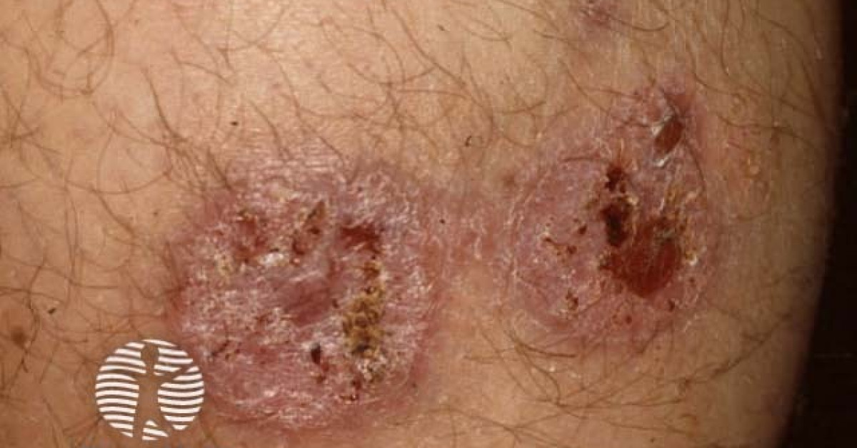
Cutaneous leishmaniasis is a sandfly-borne protozoal infection caused by various Leishmania species, presenting in UK practice as a non-healing ulcer or nodule in a returned traveller, refugee or resident of an endemic region (Mediterranean basin, Middle East, North / East Africa, Indian subcontinent, Central / South America). Travel history is essential — the lesion is frequently misdiagnosed as bacterial cellulitis, cSCC or sporotrichosis for months before correct identification. Old-world disease (L. major, L. tropica, L. infantum) is typically self-healing; new-world disease (L. braziliensis, L. mexicana) carries a risk of mucocutaneous leishmaniasis (espundia) and warrants systemic treatment. UK diagnostic and treatment guidance is via the London School of Hygiene & Tropical Medicine and specialist tropical infectious diseases services.
Epidemiology and exposure
- Sandfly vector (Phlebotomus in the old world; Lutzomyia in the new world).
- UK cases are imported — Mediterranean / Middle East / North Africa / Indian subcontinent (old world) or South / Central America (new world).
- Refugee and aid-worker / military / tourist exposure prominent.
- Incubation 1–12 weeks; lesion develops at the bite site.
- Species determines presentation, course and treatment — sample for PCR speciation.
Clinical features
- Papule → nodule → ulcer over weeks to months at site of inoculation.
- Typical lesion — single or few papulonodular ulcer with a raised indurated border ("volcano sign"); painless or mildly painful.
- Common sites — exposed skin: face, neck, dorsal hands, ankles.
- Sporotrichoid lymphocutaneous spread — secondary nodules along lymphatic drainage in some cases.
- Mucocutaneous leishmaniasis (espundia) — late mucosal involvement in L. braziliensis infection; nasopharyngeal destruction.
- Diffuse cutaneous leishmaniasis — multiple nodules in anergic patients; severe and refractory.
- Recidivans lupoid — chronic recurrence of L. tropica lesions over years.
Diagnosis
- Detailed travel history — country, region, dates, activity.
- Biopsy from active edge of the ulcer (not the necrotic centre):
- Giemsa-stained smears and tissue impression — amastigotes within macrophages.
- Tissue PCR (gold standard) with species identification.
- Culture in NNN medium — supplementary.
- Serology — limited value in cutaneous disease.
- HIV testing — particularly for diffuse / treatment-refractory disease.
- Refer to specialist tropical / infectious-diseases service (LSHTM, Royal Free, Hospital for Tropical Diseases UCLH) for species identification and treatment planning.
Management
- Decisions depend on species, lesion number, anatomical site, host immunity, and risk of mucocutaneous progression:
- Localised old-world disease, small lesion, low-risk species — watchful waiting (self-resolution over 3–12 months) or topical / intralesional therapy (cryotherapy + intralesional sodium stibogluconate, paromomycin ointment).
- Multiple / large / cosmetically sensitive / immunosuppressed — systemic therapy.
- New-world species (particularly L. braziliensis) or risk of mucocutaneous progression — systemic therapy.
- Systemic options — liposomal amphotericin B (first-line in UK), miltefosine, sodium stibogluconate (Pentostam), oral azoles (variable).
- Multidisciplinary tropical infectious-diseases input is essential — drug choice and duration are species-specific.
- Mucocutaneous disease — long-term liposomal amphotericin B; close ENT surveillance.
References
- WHO Manual for case management of cutaneous leishmaniasis in the WHO Eastern Mediterranean Region; 2014.
- Hospital for Tropical Diseases UCLH — UK leishmaniasis service.
- Aronson N et al. IDSA / ASTMH Clinical Practice Guideline for the diagnosis and treatment of leishmaniasis. Clin Infect Dis; 2017.
Spot a correction?
If any clinical statement, citation or link on this page needs updating, please email admin@skinoncology.net with the page name, the proposed correction and the supporting source.