

Cockade naevus
Cockarde naevus · concentric (target-like) naevus · cocarde naevus
A cockade (cockarde) naevus is a benign melanocytic naevus with a distinctive concentric, target-like (rosette) appearance of alternating pigmented and depigmented rings. It usually represents a benign naevus, which may be congenital or acquired, with peripheral halo-like depigmentation or central regression. Despite the unsettling clinical appearance, it is almost invariably benign. UK clinical relevance is principally to recognise the pattern and avoid unnecessary excision in young adults / adolescents.
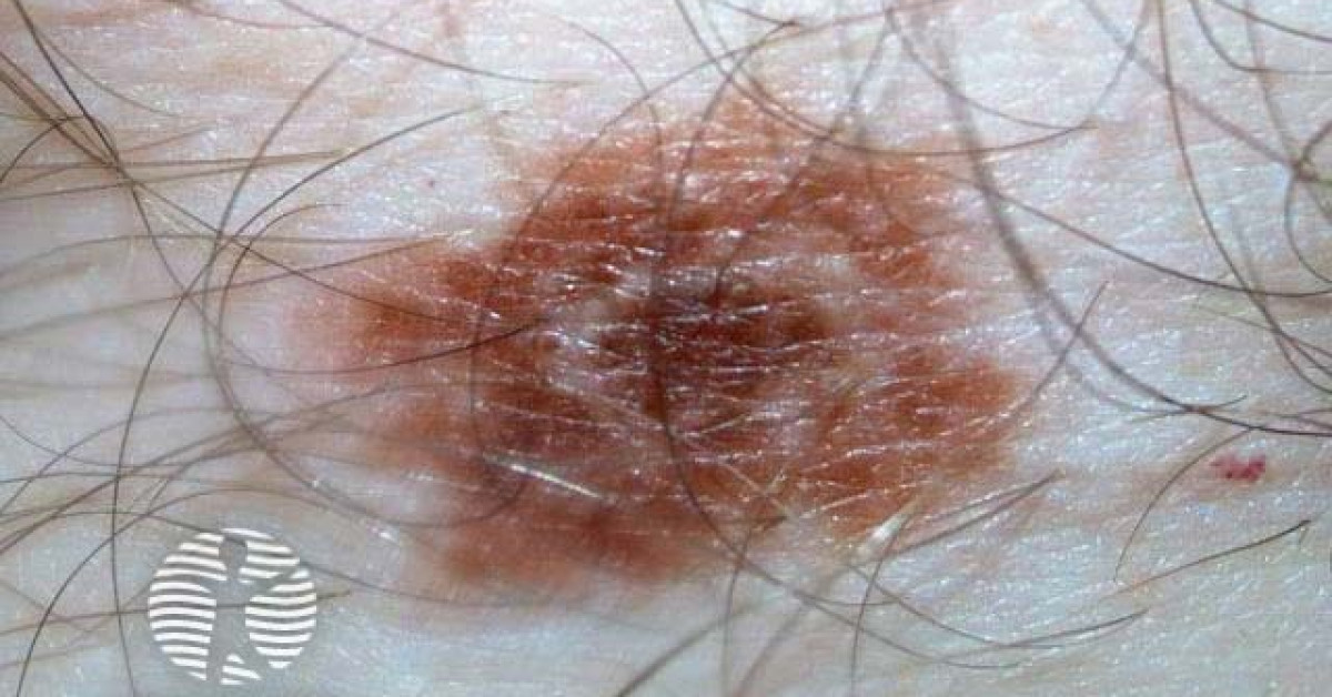
Clinical features
- Distinctive concentric ring pattern with alternating brown / tan and depigmented / pink concentric zones, like a target / cockade.
- 2-6 mm diameter typical.
- Sites: trunk, limbs.
- Adolescents / young adults commonly.
- Asymptomatic.
- Slow growth; stable over years.
- May regress spontaneously, leaving residual hyperpigmentation or hypopigmented patch.
Pathology
- Benign compound or junctional acquired melanocytic naevus.
- Concentric pigment distribution reflects variation in melanocyte / melanin density.
- Some show histological features of halo naevus — lymphocytic infiltrate in peripheral / central ring.
- No atypia; no dermal mitoses; benign Ki-67.
Dermoscopy
- Concentric rings of brown reticular network alternating with depigmented patches.
- Symmetric pattern; no atypical features.
- Cobblestone pattern of brown globules.
- No blue-white veil, no atypical pseudopods, no eccentric pigmentation.
Differentials
- Halo naevus — peripheral depigmentation; vitiligo-like halo.
- Targetoid haemosiderotic / hobnail haemangioma — vascular; biopsy.
- Erythema multiforme target lesion — acute, transient.
- Fixed drug eruption with central pigmentation.
- Combined naevus — distinct concentric components.
- Tinea corporis — annular advancing scaling edge.
- Erythema annulare centrifugum.
- Melanoma with regression — exclude with biopsy if atypia.
Management
- Clinical diagnosis with dermoscopy in classical cases.
- Reassurance — benign.
- Photographic monitoring if patient or clinician wants observation.
- Excisional biopsy if atypical features, asymmetry, growth, change in colour, irregular margin.
- Counsel on possible spontaneous regression and residual pigment change.
References
- Happle R. The Cockarde naevus: unusual variant of the common congenital melanocytic naevus. Acta Derm Venereol. 1974;54:148.
- James WD, Carter JM, Rodman OG. Pigmentary nevi (cockade, target, halo): a clinical review. Dermatol Clin. 1988;6:233-240.
- Argenziano G et al. Dermoscopy of pigmented skin lesions: results of a consensus meeting via the internet. J Am Acad Dermatol. 2003;48:679-693.
Spot a correction?
If any clinical statement, citation or link on this page needs updating, please email admin@skinoncology.net with the page name, the proposed correction and the supporting source.