Chondroid syringoma (cutaneous mixed tumour)
Cutaneous mixed tumour; benign mixed tumour of skin
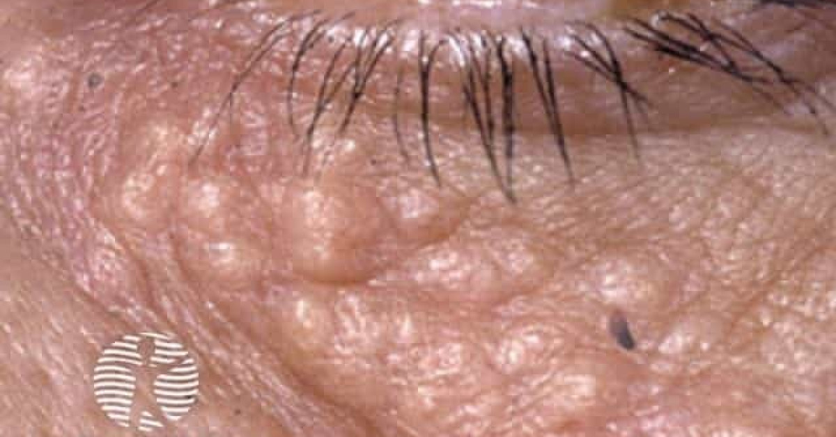
Chondroid syringoma is a benign adnexal tumour of skin showing biphasic differentiation — epithelial ductal structures within a chondromyxoid or hyalinised stroma — analogous to the pleomorphic adenoma of the salivary glands. It typically presents as a solitary, firm, slow-growing, skin-coloured nodule, 5–30 mm, on the head and neck of an adult. The diagnosis is almost always made histologically after excision because of clinical overlap with cysts, lipomas and BCC. A malignant counterpart (malignant chondroid syringoma) is rare but recognised, with metastatic potential; recurrence after incomplete excision of even benign lesions warrants careful pathology review.
Clinical features
- Solitary, firm, skin-coloured to pink nodule, 5–30 mm.
- Distribution — head and neck (commonest), upper trunk, extremities, occasionally genitalia.
- Slow growth over years; usually asymptomatic.
- Median age 40–60; slight male predominance.
- Long stability is typical of benign disease — rapid growth, ulceration or recurrence should raise concern for the malignant variant.
Histology
- Biphasic — epithelial ducts and tubules within a chondroid, myxoid or hyalinised stroma.
- Apocrine variant (predominantly head and neck) and eccrine variant (predominantly extremities) are described.
- Immunohistochemistry — epithelial cells CK7+, EMA+, often gross-cystic-disease-fluid-protein 15 (GCDFP-15) positive in apocrine variant; myoepithelial cells S100+, SMA+, p63+.
- Malignant chondroid syringoma — atypia, mitoses, infiltrative growth, necrosis; demands wide excision and staging.
Differential
- Epidermoid cyst — central punctum, cheesy contents.
- Lipoma — softer, mobile.
- BCC — pearly, telangiectatic.
- Pilomatrixoma — calcified, paediatric/adolescent.
- Apocrine adenoma; eccrine spiradenoma — overlap on biopsy.
- Salivary pleomorphic adenoma (parotid or accessory salivary tissue) — consider for periauricular lesions.
Management
- Complete surgical excision with histology — diagnostic and definitive for benign disease.
- Confirmed benign disease — reassurance, no further treatment.
- Malignant chondroid syringoma — wide local excision with margins per pathology, MDT discussion, baseline imaging (CT NCAP), surveillance.
- Recurrence after incomplete excision can occur even with benign histology — request careful re-review of margins.
References
- Hirsch P, Helwig EB. Chondroid syringoma — mixed tumor of skin, salivary gland type. Arch Dermatol; 1961.
- Mentzel T et al. Malignant chondroid syringoma — a clinicopathologic study. Histopathology; 2003.
- WHO Classification of Tumours Editorial Board. Skin Tumours, WHO Classification of Tumours, 5th ed., vol. 12. Lyon: IARC; 2025.
Spot a correction?
If any clinical statement, citation or link on this page needs updating, please email admin@skinoncology.net with the page name, the proposed correction and the supporting source.