

Cherry angioma
Campbell de Morgan spot; senile angioma; cherry haemangioma
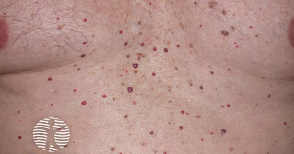
Cherry angiomas — also known as Campbell de Morgan spots — are the commonest acquired vascular lesions of adulthood, presenting as multiple bright red, dome-shaped 1–5 mm papules on the trunk and proximal limbs from the third decade onwards and progressively accumulating with age. They are entirely benign and require no treatment. Their skin-oncology relevance is twofold: (1) clinical differential from amelanotic melanoma, pyogenic granuloma, Kaposi sarcoma and angiosarcoma, particularly when atypical (large, ulcerated, atypical site, rapidly enlarging); and (2) the rare situation of "eruptive cherry angiomas" — the sudden appearance of multiple new lesions in an adult — which has been reported with some malignancies and with chemotherapy / immunotherapy adverse effects, but is usually not a stand-alone cancer marker.
Clinical features
- Bright red to violaceous, dome-shaped, smooth-surfaced papules, 1–5 mm.
- Multiple, often dozens to hundreds.
- Distribution — trunk (especially upper trunk), proximal limbs, neck; less often face.
- Onset typically third decade onwards; prevalence rises steeply with age.
- Asymptomatic; bleed minimally if traumatised.
- Variants:
- Pedunculated cherry angioma (especially trunk).
- "Multiple eruptive cherry angiomas" — sudden eruption of dozens of new lesions over weeks; reported with some malignancies, chemotherapy / immunotherapy (especially BRAF inhibitors and anti-PD-1), pregnancy, immunosuppression and after exposure to certain chemicals (mustard gas, bromides), but usually not diagnostic of occult cancer in isolation.
Dermoscopy
- Red-purple lacunae arranged in clusters ("vascular lacunae" pattern).
- Sometimes with white septae between lacunae.
- Absent — pigment network, atypical vessels, peripheral structures (which would suggest melanoma or vascular malignancy).
Differential diagnosis
- Amelanotic melanoma — particularly nodular variant; typically larger, ulcerated, growing.
- Pyogenic granuloma — solitary, friable, polypoid, rapidly growing — see monograph.
- Kaposi sarcoma — multiple violaceous patches / plaques; HIV / iatrogenic / classical context — see monograph.
- Cutaneous angiosarcoma — older patient; head / scalp; bruise-like — see monograph.
- Cutaneous metastasis — particularly from breast, RCC; firmer, growing.
- Spider angioma — central vessel with radiating telangiectasias; face, upper trunk; chronic liver disease.
- Angiokeratoma — darker red-purple, hyperkeratotic, on scrotum / vulva (Fordyce) or extremities (Mibelli); see monograph.
- Biopsy any atypical or solitary "cherry angioma" — especially if large, ulcerated, in an unusual location or rapidly growing.
Management
- Reassurance — no treatment required for typical asymptomatic lesions.
- Cosmetic / haemostatic options:
- Electrosurgery (needle / ball cautery).
- Pulsed dye laser (585 / 595 nm).
- Long-pulsed Nd:YAG laser (1064 nm).
- Intense pulsed light.
- Shave excision for very large lesions.
- Cryotherapy.
- Eruptive cherry angiomas — review systemic context (drug history, recent malignancy, pregnancy); consider full skin examination and age-appropriate cancer screening; biopsy any atypical lesion; reassure if no underlying cause found.
References
- Kim JH et al. Eruptive cherry angiomas — review. Ann Dermatol; 2013.
- Borghi A et al. Eruptive cherry angiomas associated with vemurafenib treatment. Acta Derm Venereol; 2017.
Spot a correction?
If any clinical statement, citation or link on this page needs updating, please email admin@skinoncology.net with the page name, the proposed correction and the supporting source.