Adenoid cystic carcinoma of skin
Primary cutaneous adenoid cystic carcinoma (PCACC); cribriform sweat-gland carcinoma
Primary cutaneous adenoid cystic carcinoma is a rare, slow-growing, infiltrative adnexal carcinoma with eccrine differentiation, sharing the same characteristic cribriform / tubular / solid architecture and propensity for perineural invasion seen in salivary-gland adenoid cystic carcinoma. The recurrent MYB-NFIB gene fusion (t(6;9)) found in salivary ACC is also present in many cutaneous cases, supporting a true biological relationship. The scalp is the commonest site, followed by face, trunk and extremities, in middle-aged to elderly adults. Despite a deceptively indolent clinical course, perineural extension along cranial nerves can be extensive and clinically silent, making complete surgical clearance difficult and local recurrence common (50–70% with conventional excision). Mohs micrographic surgery substantially reduces recurrence. Distant metastasis is rare but late.
Clinical features
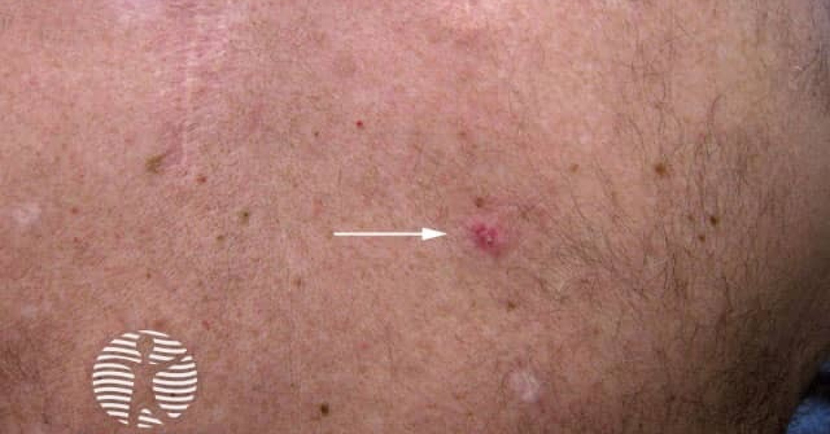
- Slowly enlarging, ill-defined, indurated dermal/subcutaneous nodule or plaque.
- Most common site: scalp (~50%), then face, trunk, extremities.
- Median age 60; M:F roughly equal.
- Often present for years to decades before diagnosis.
- Symptoms of perineural extension — paraesthesia, dysaesthesia, cranial-nerve palsy — should raise suspicion.
- Differential: BCC (especially morphoeic), microcystic adnexal carcinoma, primary cutaneous mucinous carcinoma, metastatic salivary ACC.
Histology & molecular
- Three classic architectural patterns — usually mixed:
- Cribriform — sieve-like nests with pseudoglandular spaces filled with PAS-positive basement-membrane material.
- Tubular — small glandular structures.
- Solid — sheets of basaloid cells; higher grade and more aggressive.
- Two cell populations: small basaloid epithelial cells and surrounding myoepithelial cells.
- Marked perineural invasion in >50% — often extends far beyond the clinical lesion.
- Immunohistochemistry: CK7+, p63+ (myoepithelial), CD117 (KIT)+, EMA+; MYB protein nuclear positive.
- MYB-NFIB fusion (t(6;9)) in 50–80% — supports primary cutaneous origin and aligns with salivary ACC.
- Differential vs metastatic salivary ACC requires correlation with head/neck and chest imaging.
Management
- Mohs micrographic surgery — first-line; reduces local recurrence to ~5–20% (compared with 50–70% for conventional excision) by mapping the deep and peripheral perineural extension.
- Wide local excision with at least 1–2 cm margins and complete histological assessment of the entire deep margin (peripheral en-face frozen section / "slow Mohs") if Mohs is unavailable.
- MRI of head/neck if perineural invasion is histologically demonstrated — to assess proximal extent and skull-base.
- Adjuvant radiotherapy for incomplete margins, named-nerve perineural invasion or recurrent disease.
- Sentinel lymph node biopsy not routinely performed; nodal metastasis uncommon.
- Systemic therapy for advanced / metastatic disease — limited evidence; multikinase inhibitors (lenvatinib), MYB-targeted therapies in development.
Prognosis
Local recurrence is the main long-term concern (50–70% with conventional excision; substantially lower with Mohs). Distant metastasis <10% but late, with lung, bone and CNS most common sites. Long-term surveillance for at least 10–15 years is appropriate, with low threshold for re-imaging if perineural symptoms develop.
References
- Krunic AL et al. Primary cutaneous adenoid cystic carcinoma — comprehensive review. J Am Acad Dermatol; 2003.
- North JP et al. MYB-NFIB rearrangement in primary cutaneous adenoid cystic carcinoma. Mod Pathol; 2018.
Spot a correction?
If any clinical statement, citation or link on this page needs updating, please email admin@skinoncology.net with the page name, the proposed correction and the supporting source.