Acral melanocytic naevi
Acral lentiginous naevus (benign); palmoplantar naevus; volar naevus
Acral melanocytic naevi are benign pigmented lesions on palms, soles and digits. They are far more conspicuous in Fitzpatrick IV–VI skin, where they make up a substantial proportion of all naevi, and remain the principal benign differential for acral lentiginous melanoma. Three benign dermoscopic patterns predominate — parallel-furrow, lattice-like and fibrillar — all of which reflect pigment in the narrower furrows of the dermatoglyphics rather than the wider ridges. The parallel-ridge pattern of acral lentiginous melanoma reverses this distribution and is the single most useful early warning sign on dermoscopy.
Clinical features
- Well-circumscribed, evenly pigmented brown macule or thin papule on palms, soles, fingers or toes.
- Usually 2–8 mm; larger lesions warrant closer scrutiny.
- Common from childhood; persistence into adulthood is normal.
- Proportionally commoner in Fitzpatrick IV–VI skin.
- Subungual / nail-matrix naevi present as longitudinal melanonychia, typically < 3 mm wide with uniform colour — see subungual melanoma for melanoma red flags.
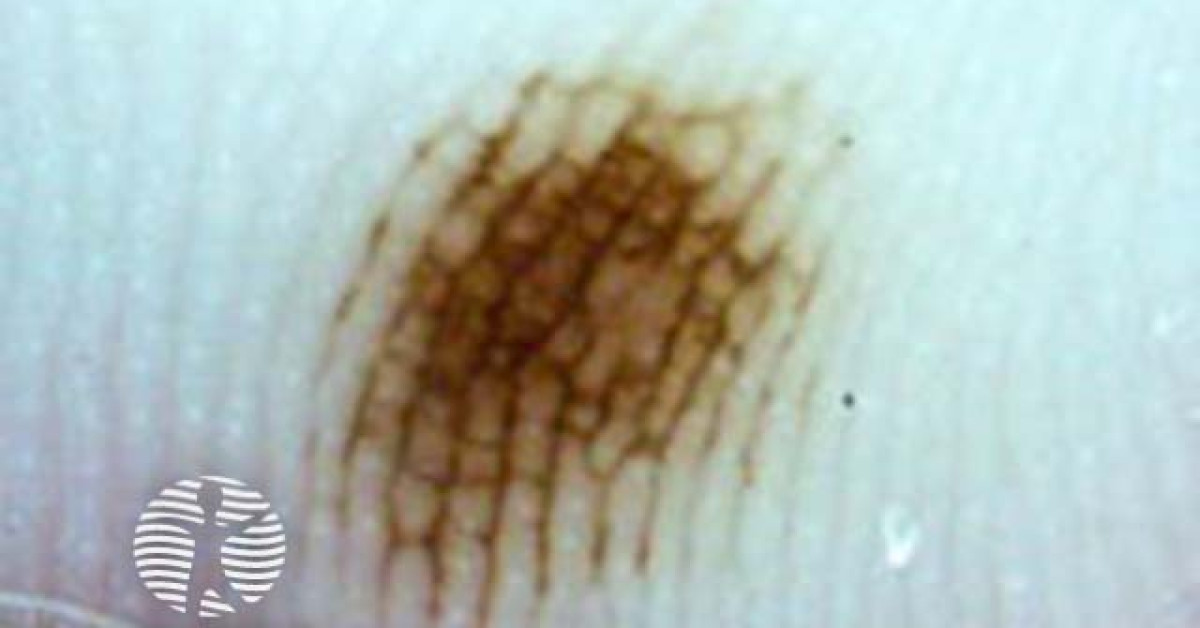
Benign dermoscopic patterns
- Parallel-furrow pattern — pigment along the narrower furrows of the dermatoglyphics; commonest benign pattern, particularly on the sole.
- Lattice-like pattern — pigment crossing the furrows with additional perpendicular lines; common on the arch.
- Fibrillar pattern — fine oblique pigmented lines crossing the surface; common on weight-bearing areas.
- Less common but benign — globular, reticular, homogeneous patterns (mostly in children and on the dorsum / less acral areas).
Parallel-ridge — the melanoma warning
- The parallel-ridge pattern — pigment deposited along the wider ridges rather than the narrower furrows — is the classical dermoscopic finding of acral lentiginous melanoma.
- High-specificity sign in trained hands; the original Saida series reported sensitivity in the mid-80% range and specificity ~99% for early acral melanoma, but performance depends on training and lesion mix.
- Any acral lesion with parallel-ridge pattern, multicomponent pattern, asymmetric pigmentation, blue-white veil or peripheral pigment must be referred urgently.
- The CASH (Colour, Architecture, Symmetry, Homogeneity) approach helps risk-stratify uncertain acral lesions.
Special situations
- Acral naevi in skin of colour — a major equity issue. ALM is over-represented in Fitzpatrick IV–VI and frequently presents late; threshold for biopsy should be lower in this population.
- Subungual naevus vs subungual melanoma — Hutchinson sign (pigment extending onto the nail fold) is concerning but not pathognomonic (pseudo-Hutchinson exists in benign disease).
- Children and adolescents — congenital and acquired acral naevi are common; the parallel-ridge pattern is seen far less reliably as a melanoma marker in this group, and benign acral naevi may show transient atypical patterns.
Management and biopsy
- Routine review for stable, symmetrical lesions with benign dermoscopic patterns.
- Biopsy and refer if asymmetry, parallel-ridge, multicomponent or evolving features.
- Excisional biopsy preferred over shave for acral pigmented lesions where melanoma is possible — preserves depth.
- Nail-matrix biopsy for atypical longitudinal melanonychia > 3 mm wide, asymmetric, or with Hutchinson sign.
References
- Saida T et al. Significance of dermoscopic patterns in detecting early acral lentiginous melanoma. J Am Acad Dermatol; 2004.
- Phan A et al. Acral lentiginous melanoma — clinicoprognostic features in a cohort. Br J Dermatol; 2010.
- Bristow IR, de Berker DA. Acral lentiginous melanoma — clinical update. Br J Dermatol; 2018.
- NICE NG14. Melanoma: assessment and management. London: NICE; 2015 (last updated 27 July 2022).
Spot a correction?
If any clinical statement, citation or link on this page needs updating, please email admin@skinoncology.net with the page name, the proposed correction and the supporting source.