Acral lentiginous melanoma
ALM
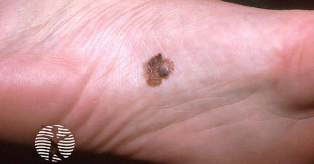
Acral lentiginous melanoma is a histological subtype of melanoma arising on glabrous (non-hair-bearing) palmar, plantar and subungual skin. Although it accounts for only ~5% of melanomas in white populations, it is the most common cutaneous melanoma subtype in people of colour (Asian, African, Hispanic). Stage-for-stage prognosis is similar to other melanomas, but presentation is frequently delayed, leading to worse overall outcomes. Driver mutations differ — KIT and CCND1 amplifications are over-represented; BRAF V600E is uncommon (~10–15%).
Epidemiology
- ~2–5% of all cutaneous melanomas in white populations.
- Up to ~30–50% of melanomas in people of African, Asian or Hispanic descent (proportions vary widely across cohorts; older series quoted higher figures).
- Median age 60–70; equal sex distribution.
- UV is not a major causative factor — incidence is broadly similar across populations regardless of latitude.
Clinical features
- Plantar: commonest site (~half). Slow-growing irregular pigmented patch on weight-bearing or non-weight-bearing sole. Often misdiagnosed as haematoma, callus or fungal nail.
- Palmar: less common; broad pigmented macule/patch.
- Subungual: longitudinal melanonychia >3 mm wide, irregular pigmentation, Hutchinson's sign (periungual extension).
- ABCDE criteria apply but with a lower threshold; for nail, use ABCDEF (Age, Brown/Black band >3 mm, Change, Digit (thumb > great toe), Extension to nailfold, Family/personal history) or CUBED (Coloured lesion, Uncertain diagnosis, Bleeding, Enlargement, Delay in healing).
Dermoscopy
- Parallel ridge pattern — pigment along the ridges (raised lines) of dermatoglyphics — highly specific for melanoma on volar skin.
- Parallel furrow pattern, lattice-like, fibrillar pattern — usually benign acral naevus.
- Diffuse irregular pigmentation, multicomponent pattern — suspicious.
- Subungual: longitudinal lines of irregular colour, width and spacing; granular pigment in proximal nail fold.
Diagnosis
- Excisional biopsy preferred when feasible (incisional or punch acceptable for very large or anatomically constrained sites).
- For subungual lesions: longitudinal nail matrix biopsy (3 mm punch, lateral matrix shave, or full longitudinal excision) to capture matrix epithelium.
- Histology: lentiginous proliferation of atypical melanocytes along the dermo-epidermal junction with later dermal invasion.
Management
- Wide local excision per NICE NG14 stage-based margins: stage 0 at least 0.5 cm; stage I 1 cm; stage II 2 cm, using 1 cm only if a 2 cm margin would cause unacceptable disfigurement or morbidity.
- Sentinel lymph node biopsy considered according to NICE NG14 §1.4.3–1.4.4.
- Subungual: traditionally distal amputation at the DIPJ (toe) or IPJ (thumb). For subungual melanoma in situ, digit-preserving nail-unit excision with full-thickness skin graft reconstruction can be considered in specialist hands. Invasive subungual melanoma should be planned through the melanoma MDT, with oncological clearance taking priority over reconstruction.
- BRAF mutation is uncommon in ALM; KIT testing useful in advanced disease — selected patients respond to imatinib.
- Anti-PD-1 (pembrolizumab, nivolumab) — overall response rates lower than for cutaneous melanoma but still meaningful and remain first-line for advanced disease.
Prognosis
Stage-for-stage outcomes are broadly similar to non-acral cutaneous melanoma. However, ALM commonly presents at higher Breslow thickness because of delayed diagnosis (mistaken for benign lesions, plantar callus, fungal nail or haematoma) and lack of awareness in skin of colour — so overall survival is worse. Public and clinician education to think melanoma in dark-skinned patients with foot or nail lesions is crucial.
References
- Bradford PT et al. Acral lentiginous melanoma: incidence and survival patterns in the United States, 1986–2005. Arch Dermatol; 2009.
- Saida T et al. Significance of dermoscopic patterns in detecting malignant melanoma on acral volar skin. Arch Dermatol; 2004.
- Cochran AM et al. Subungual melanoma: a review of current treatment. Plast Reconstr Surg; 2014.
Spot a correction?
If any clinical statement, citation or link on this page needs updating, please email admin@skinoncology.net with the page name, the proposed correction and the supporting source.