InfectionYeastICD-10 B36.0
Pityriasis versicolor
Tinea versicolor · Malassezia versicolor infection
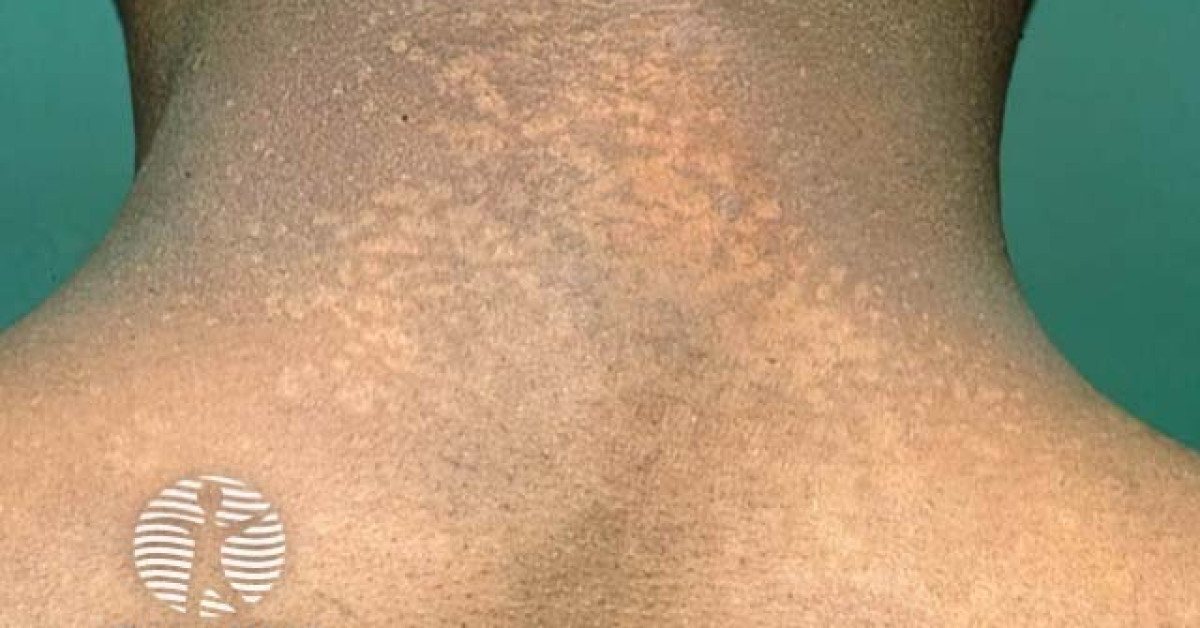
Pityriasis versicolor is a superficial cutaneous mycosis caused by overgrowth of skin commensal Malassezia spp. (formerly Pityrosporum). The yeasts switch to a hyphal form on lipid-rich skin, producing dyspigmented scaly macules on the trunk, upper arms and neck. It is among the most common DDx for hypo- or hyperpigmented patches in young adults and frequently mimics tinea, post-inflammatory dyspigmentation, vitiligo and early hypopigmented mycosis fungoides.
CurrentLast reviewed 16 May 2026
Microbiology
- Lipophilic commensal yeasts: Malassezia globosa (commonest), M. furfur, M. sympodialis, M. restricta.
- Conversion from yeast → hyphal phase on lipid-rich skin (sebaceous areas) drives disease.
- Triggers: humid warm climate / season, sweating, occlusive clothing, oily skin, oral contraceptives, immunosuppression (including ICI, ciclosporin), pregnancy.
- Azelaic acid produced by Malassezia inhibits tyrosinase → hypopigmentation in tanned skin.
Clinical features
- Multiple small (5-10 mm) coalescing macules; well-demarcated.
- Variable colour:
- Hypopigmented on tanned / Fitzpatrick III-VI skin.
- Hyperpigmented / pinkish-tan on pale skin.
- Erythematous in inflammatory phase.
- Fine bran-like (furfuraceous) scale on stretching the skin ("Besnier sign").
- Sites: upper chest, upper back, shoulders, neck, proximal arms; rarely face.
- Chronic, recurrent; warm-weather flares.
- Pruritus mild or absent.
Investigations
- Clinical diagnosis usually sufficient.
- Wood lamp: golden-yellow fluorescence in ~30%.
- KOH microscopy: short hyphae with clusters of round yeasts ("spaghetti and meatballs").
- Dermoscopy: thin scale within follicular ostia.
- Reserve skin biopsy for atypical or refractory cases — PAS shows yeast / hyphae.
Differentials
- Vitiligo — complete depigmentation, no scale, Wood lamp accentuates.
- Hypopigmented mycosis fungoides — chronic, atrophic, in children / young adults of darker skin; biopsy.
- Pityriasis alba — facial in children with atopic eczema.
- Post-inflammatory hypopigmentation.
- Tinea corporis — annular advancing edge.
- Confluent and reticulated papillomatosis (Gougerot-Carteaud) — reticulate, intermammary.
- Seborrhoeic dermatitis overlaps; Malassezia involved.
Management
- Topical (first-line):
- Ketoconazole 2% shampoo applied to wet skin, left 5-10 minutes, rinsed; daily for 5-7 days.
- Selenium sulfide 2.5% lotion / shampoo.
- Zinc pyrithione shampoo.
- Topical azoles (clotrimazole, miconazole, ketoconazole cream) for limited disease.
- Oral (extensive / refractory / recurrent):
- Itraconazole 200 mg OD for 7 days.
- Fluconazole 300 mg weekly for 2-4 weeks.
- Avoid oral ketoconazole (hepatotoxicity).
- Counsel:
- Pigment change persists for weeks-months after fungal eradication (does not equal treatment failure).
- Recurrence common (~60% at 2 years) — prophylactic ketoconazole shampoo monthly may help.
References
- Hald M et al. Evidence-based Danish guidelines for the treatment of Malassezia-related skin diseases. Acta Derm Venereol. 2015;95:12-19.
- Crespo Erchiga V, Florencio VD. Malassezia yeasts and pityriasis versicolor. Curr Opin Infect Dis. 2006;19:139-147.
- Gupta AK, Foley KA. Antifungal treatment for pityriasis versicolor. J Fungi (Basel). 2015;1:13-29.
- British Association of Dermatologists. Pityriasis versicolor — patient information leaflet. London: BAD; 2023.
Spot a correction?
If any clinical statement, citation or link on this page needs updating, please email admin@skinoncology.net with the page name, the proposed correction and the supporting source.