Mucocele
Mucous extravasation phenomenon; mucous retention cyst; ranula (when on floor of mouth)
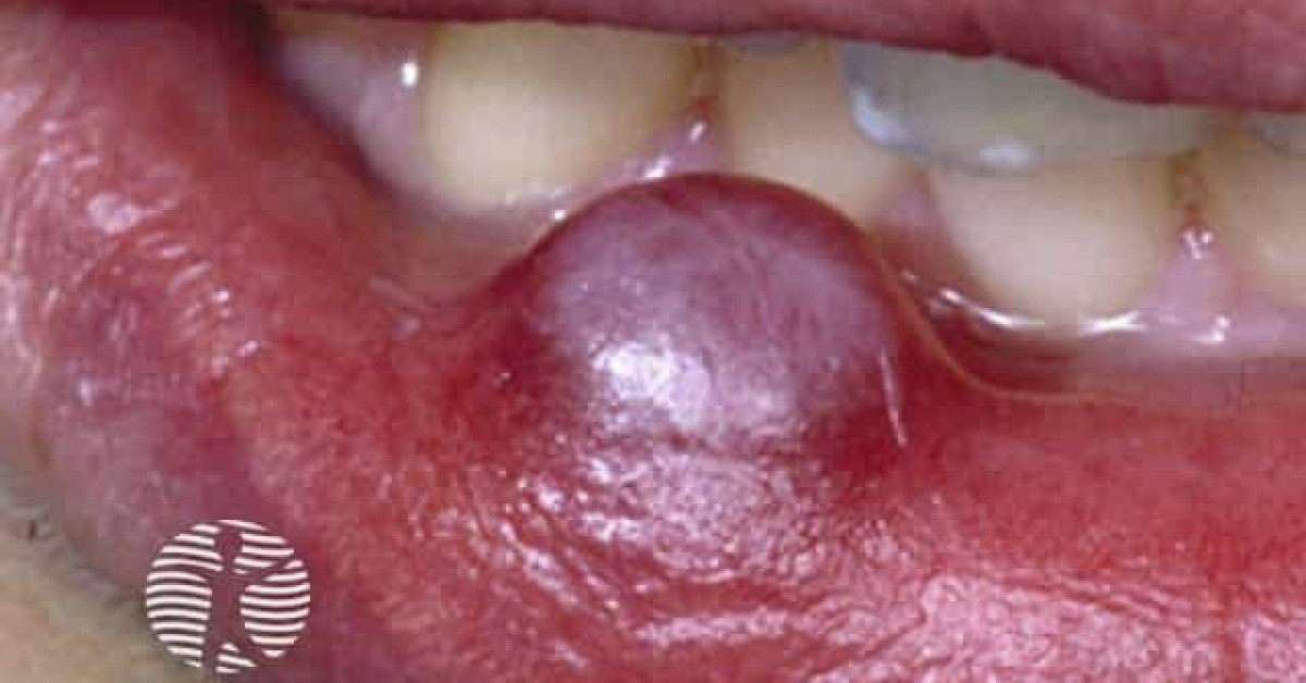
A mucocele is a common benign lesion of the oral mucosa caused by extravasation of mucus from a minor salivary gland into the surrounding stroma — typically following minor lip-bite trauma. It presents as a dome-shaped translucent blue or skin-coloured fluctuant swelling, most commonly on the inner lower lip, and is a recurrent reason for reassurance in primary care and dental practice. A larger variant on the floor of the mouth is known as a ranula. Mucocele is most often confused with venous lake, blood blister, fibroma, salivary gland tumour or mucinous carcinoma — but the typical recurrent fluctuant nature in a patient with lip-biting habit is diagnostic. Excision or marsupialisation cures most cases.
Clinical features
- Dome-shaped, fluctuant, translucent blue or skin-coloured swelling, 2–10 mm, on the inner lower lip.
- Frequently spontaneously ruptures and recurs.
- Other sites — buccal mucosa, tongue (ventral), floor of mouth (ranula), occasionally palate.
- Patient often reports trauma — lip biting, dental procedure, mucosal piercing.
- Asymptomatic except for fluctuant feel; may interfere with eating / speech for larger lesions.
- Sublingual ranula presents as a larger blue swelling on the floor of the mouth; "plunging" ranula extends into the neck.
Differential
- Venous lake — older patient, on lip vermilion (not inner mucosa), compressible, dark blue.
- Salivary gland tumour (e.g. pleomorphic adenoma) — firm, fixed, slow-growing, not fluctuant.
- Mucinous carcinoma — firm, infiltrative, atypical features.
- Fibroma / fibrous polyp — firm, white-grey, non-fluctuant.
- Blood blister — traumatic; recent onset; resolves spontaneously.
- Lymphangioma — paediatric; cystic; may have surface vesicles.
Histology
- Pseudocyst — no true epithelial lining; cavity contains mucin and inflammatory cells.
- Surrounding granulation tissue, foamy macrophages, occasional multinucleated giant cells.
- Adjacent residual minor salivary-gland duct often visible.
- Distinction from true mucinous tumours is histologically straightforward.
Management
- Some lesions resolve spontaneously over weeks to months — observation is reasonable for an asymptomatic recent-onset mucocele.
- Recurrent or persistent lesions — surgical excision (including the underlying minor salivary glands) is curative; performed under local anaesthesia.
- Marsupialisation — alternative for larger or sublingual lesions.
- CO₂ laser ablation or cryotherapy in selected cases.
- Ranula — sublingual / plunging variants — refer to oral surgery; sublingual gland excision often required.
- Counsel patient about lip-biting habit modification to prevent recurrence.
References
- Bagán JV et al. Mucocele of the salivary gland — clinical and epidemiological study. Med Oral Patol Oral Cir Bucal; 2008.
- Speight PM, Barrett AW. Oral mucoceles. Periodontol 2000; 2009.
Spot a correction?
If any clinical statement, citation or link on this page needs updating, please email admin@skinoncology.net with the page name, the proposed correction and the supporting source.