Merkel cell carcinoma
Also known as: MCC; trabecular carcinoma
Merkel cell carcinoma is a rare, aggressive cutaneous neuroendocrine tumour with significant metastatic potential. Around 70–80% of cases in contemporary series are driven by Merkel cell polyomavirus; the remainder are UV-induced. Multimodality management is essential.
Clinical features
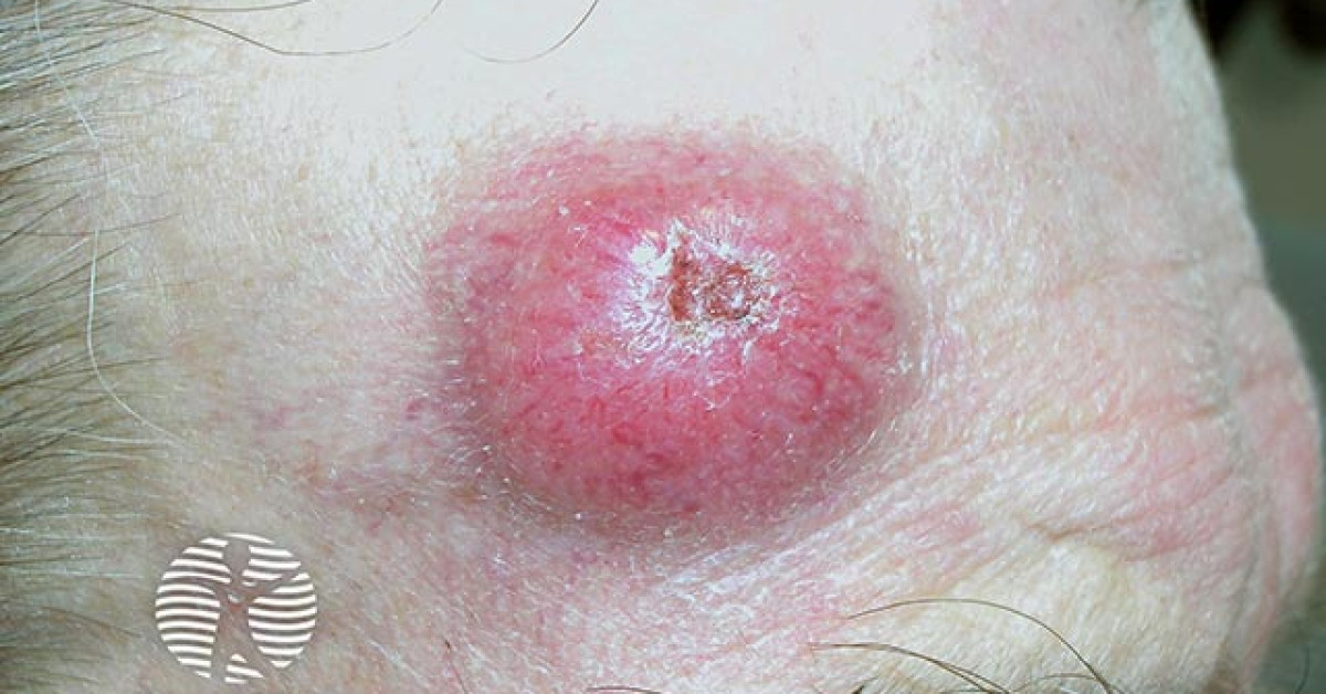
MCC typically presents as a rapidly enlarging, painless, firm, red or violaceous nodule on sun-exposed skin of older patients. Head and neck (~50%) and extremities most common. Ulceration is unusual at presentation.
AEIOU risk features
- Asymptomatic
- Expanding rapidly (weeks–months)
- Immunosuppressed
- Older than 50
- UV-exposed / fair skin
Three or more features warrant urgent biopsy.
Diagnosis
Histology required. Immunohistochemistry is pathognomonic:
- CK20 positive with paranuclear dot-like pattern.
- Neuroendocrine markers: synaptophysin, chromogranin, CD56 positive.
- TTF-1 negative — essential to exclude metastatic small-cell lung cancer.
Baseline staging imaging is routine — PET-CT preferred where available, with MRI brain if symptomatic.
Management
Localised disease
- Guideline Wide local excision with 1–2 cm margin to fascia is the standard. Mohs has a niche role at selected facial sites where tissue conservation matters, performed at specialist centres with appropriate frozen-section / IHC interpretation and without compromising SLNB workflow — it is not the routine standard.
- Local MDT SLNB for clinically node-negative (~30% positive).
- Consensus Adjuvant radiotherapy to primary site (reduces local recurrence ~3× in most series).
- Consensus Adjuvant nodal radiotherapy for SLN-positive where completion dissection is declined.
Metastatic disease
- NICE TA Avelumab — NICE TA691, first-line.
- Trial Pembrolizumab — alternative PD-1.
- Consensus Platinum-etoposide — rapid short-lived responses; reserve for ICI-refractory.
Every MCC case should be discussed at the local specialist skin cancer MDT with input from dermatology, plastic surgery, clinical oncology and medical oncology given rarity and treatment complexity.
Follow-up
- After radical treatment: clinical examination every 3–6 months for the first 3 years, then every 6 months until year 5.
- After 5 years: annual lifelong general physical examination including complete skin check.
- Each review should include full-skin examination, scar / in-transit field assessment and regional lymph-node examination.
- Cross-sectional imaging may be proposed for higher-risk patients, but should be MDT-led rather than a fixed interval for everyone.
References
- Gauci ML et al. EADO/EORTC/UNITE. Diagnosis and treatment of Merkel cell carcinoma: European consensus-based guidelines. Eur J Cancer; 2022.
- NICE TA691. Avelumab for untreated metastatic Merkel cell carcinoma. London: NICE; 2021.
- Nghiem PT et al. Three-year survival, correlates and salvage therapies with first-line pembrolizumab for advanced MCC. J Immunother Cancer; 2021.
Spot a correction?
If any clinical statement, citation or link on this page needs updating, please email admin@skinoncology.net with the page name, the proposed correction and the supporting source.