Glomus tumour
Solitary glomus tumour; glomangioma (vascular variant); glomangiomyoma; "subungual glomus tumour" (the most common clinical presentation)
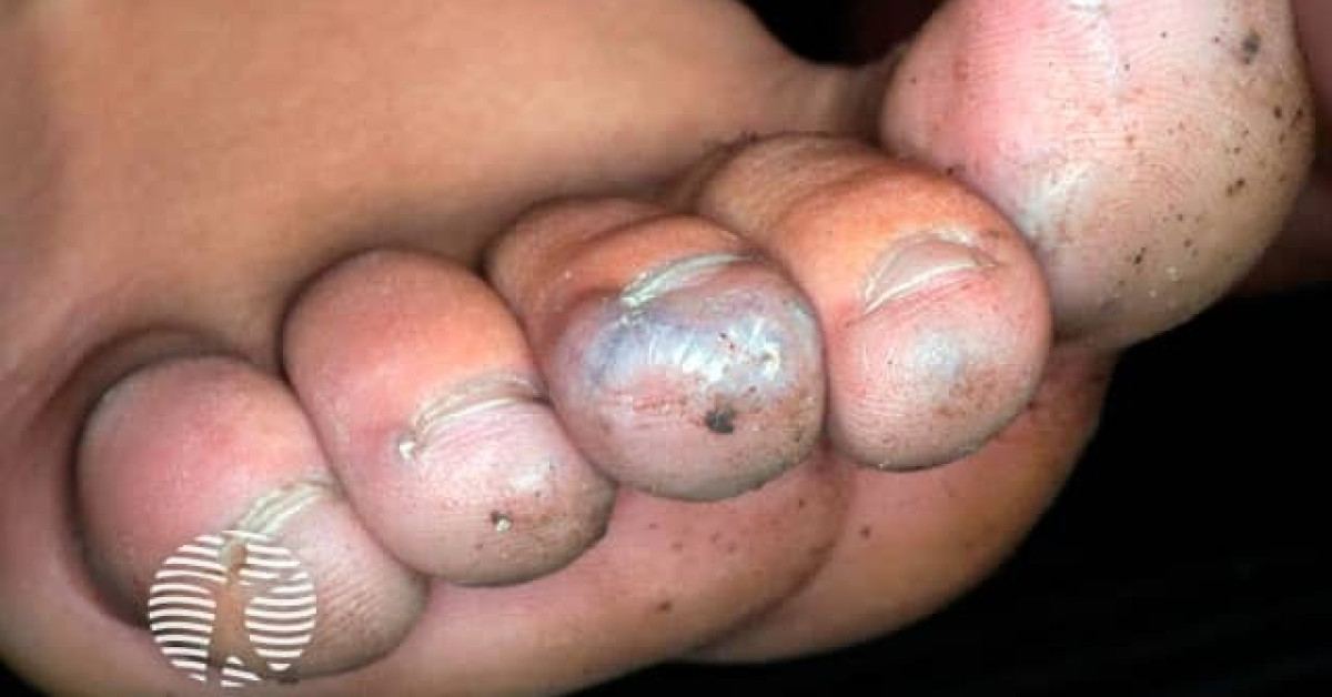
A glomus tumour is an uncommon, benign, painful neoplasm derived from the modified smooth-muscle cells of the cutaneous arteriovenous shunt — the glomus body — which regulates blood flow and temperature in acral skin. The classic presentation is a 2–10 mm bluish-red subungual nodule causing exquisite paroxysmal pain, dramatic sensitivity to cold and pinpoint pressure tenderness — confirmed at the bedside by the Love test (pin-pressure with a paperclip reproduces sharp pain) and the Hildreth test (pain abolished by limb-tourniquet inflation). MRI of the digit is the gold-standard imaging investigation. Surgical excision through a lateral or transungual approach is curative. Rare malignant variants — glomangiosarcoma — exist but represent <1% of glomus tumours.
Clinical features — the classical triad
- 1. Spontaneous and paroxysmal pain — sharp, throbbing, often nocturnal, sometimes excruciating.
- 2. Cold sensitivity — pain provoked dramatically by exposure to cold (running cold water, cold drinks, cold air).
- 3. Pinpoint tenderness — focal pain on light pressure with a pen tip / paperclip ("Love sign").
- Onset — gradual over months to years; often years of misdiagnosis as ingrowing nail, paronychia, neuroma or psychogenic pain.
- Median age 30–50; F>M.
- Visible blue-red dot or longitudinal ridge under the nail in many cases; subungual erythronychia (red longitudinal nail line) is a classic clue.
- Distal nail-bed concavity from pressure on the underlying bone in long-standing lesions.
- Multiple glomus tumours — uncommon; consider familial / hereditary multiple glomus tumours (autosomal dominant GLMN gene mutations).
- Location — >75% subungual (especially fingers); also volar finger pulp, hand, foot, and rare extracutaneous sites (stomach, lung, mediastinum).
Bedside diagnostic tests
- Love test — paperclip / pen tip pressure on the suspected lesion reproduces sharp severe pain.
- Hildreth test — pain abolished by inflating a tourniquet around the limb (proximal exsanguination), and reproduced on tourniquet release.
- Cold sensitivity test — pain reproduced by immersing the digit in cold water or applying ice.
- Sensitivity 80–100%, specificity 90–100% when all three positive.
Imaging
- MRI of the digit — gold standard; small (mm-scale) well-defined lesion in the nail bed, hyperintense on T2-weighted imaging with avid contrast enhancement.
- Ultrasound — increasingly used; well-defined hypoechoic nodule with prominent vascular flow on Doppler.
- Plain X-ray — bone erosion / scalloping in 30–50%; not diagnostic but supportive.
Management
- Surgical excision is curative.
- Approach options:
- Lateral / periungual approach — preserves the nail apparatus; preferred when the lesion is laterally located.
- Trans-ungual approach (nail-plate avulsion + matrix incision + tumour excision + matrix repair + nail-plate replacement) — for centrally located lesions.
- Refer to a hand surgeon for nail-unit-preserving surgery to optimise functional and cosmetic outcome.
- Symptom relief usually immediate; recurrence ~10% — usually due to incomplete excision.
- Histology — confirms diagnosis and excludes the rare glomangiosarcoma.
Malignant glomus tumour (glomangiosarcoma) — rare
- Criteria (Folpe et al., 2001) — any one of: deep location and size >2 cm; atypical mitotic figures; marked nuclear atypia and mitotic count >5/50 HPF.
- Behaviour — locally aggressive with risk of metastasis; manage as soft-tissue sarcoma.
- Wide local excision + sarcoma MDT.
References
- Mravic M et al. Clinical management of glomus tumors of the hand. Hand (NY); 2015.
- Folpe AL et al. Atypical and malignant glomus tumors — analysis of 52 cases. Am J Surg Pathol; 2001.
Spot a correction?
If any clinical statement, citation or link on this page needs updating, please email admin@skinoncology.net with the page name, the proposed correction and the supporting source.