

Fixed drug eruption
FDE · generalised bullous FDE (GBFDE) when extensive
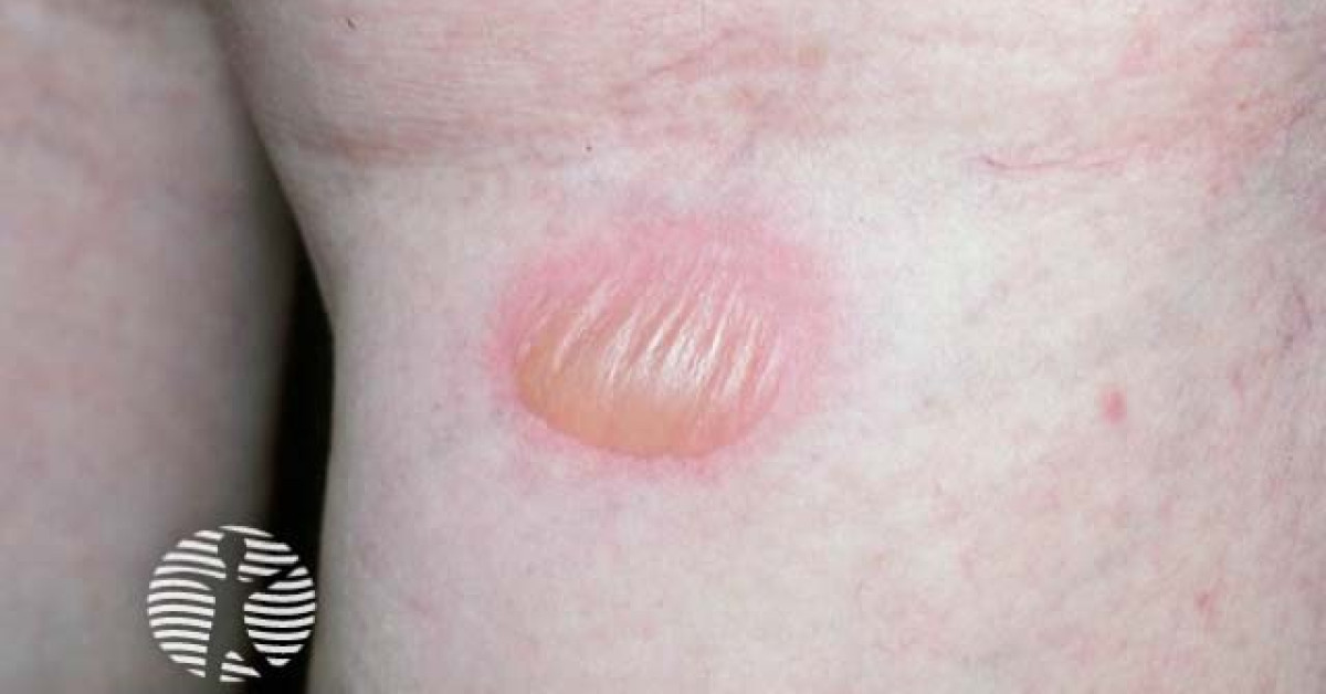
Fixed drug eruption is a delayed-type hypersensitivity reaction characterised by single or few round, sharply demarcated, dusky red-violet patches that recur at the same site with every re-exposure to the culprit drug. Common offenders are paracetamol, sulfonamides, NSAIDs, tetracyclines, antibiotics and laxatives (phenolphthalein historically). Residual hyperpigmented patches commonly persist for months and are a frequent skin-oncology DDx for naevus or melanocytic neoplasia. The generalised bullous variant (GBFDE) can overlap clinically with SJS/TEN.
Aetiology
- Common culprits:
- Paracetamol, NSAIDs (ibuprofen, mefenamic acid, naproxen).
- Sulfonamides (sulfamethoxazole, sulfasalazine).
- Antibiotics — tetracyclines, fluoroquinolones, β-lactams.
- Anticonvulsants — carbamazepine, phenytoin.
- Laxatives — phenolphthalein (historical).
- Allopurinol, antimalarials, OCP.
- Mechanism: CD8+ memory T cells resident in the epidermis at the previously affected site re-activate on drug exposure.
- Onset minutes to days after re-exposure; faster with each recurrence.
Clinical features
- One or few round / oval sharply demarcated dusky red-violet patches; 1-10 cm; well-circumscribed.
- Sites: lips, genitalia (penis, glans), face, hands, mucosae; recur at the same site with each re-exposure.
- Burning / itching with new lesions; bulla may form in centre.
- Resolution over 2-4 weeks with characteristic residual hyperpigmentation that may persist for months / years.
- Generalised bullous fixed drug eruption (GBFDE): multiple large dusky patches with bullae; can affect >10% BSA; potentially severe and confused with SJS/TEN — mucosal involvement is typically minimal.
- Variants: non-pigmenting, bullous, urticarial, eczematous, mucosal-only (oral / genital), psoriasiform.
Investigations
- Clinical diagnosis based on history + same-site recurrence.
- Drug provocation testing: oral / topical / patch testing at the affected site can confirm culprit (specialist setting).
- Skin biopsy of active lesion: interface dermatitis, vacuolar degeneration, dyskeratotic keratinocytes, dermal melanophages (pigment-incontinence — accounts for residual hyperpigmentation).
- Detailed drug history (including OTC, herbal, intermittent — paracetamol commonly missed).
Differentials
- Naevus / melanocytic lesion — single round pigmented patch on glans / lip; biopsy if uncertain.
- Erythema multiforme — true target lesions; HSV-driven.
- Phytophotodermatitis — streaky pattern; phototoxic.
- Genital herpes — vesicles; PCR.
- Fixed pigment lesion — lichen planus, sarcoidosis.
- SJS / TEN / GBFDE — generalised; mucosal involvement.
- Tinea, candidiasis in genital cases.
Management
- Withdraw and avoid culprit drug — pharmacy alert, MedicAlert.
- Symptomatic:
- Mid-to-potent topical corticosteroids during active phase.
- Oral antihistamines for pruritus.
- Cool compresses for bullous lesions.
- GBFDE:
- Admit if extensive bullae, systemic features, or diagnostic uncertainty (SJS/TEN exclusion).
- Supportive care; systemic corticosteroids in selected severe cases.
- Residual hyperpigmentation:
- Sun protection; hydroquinone 4%; tretinoin; Q-switched / picosecond laser if persistent.
- Counsel: pigment may take months-years to fade; persistence common, particularly in Fitzpatrick IV-VI.
- Document drug allergy and counsel re class cross-reactivity (e.g. sulfonamide antibiotics vs sulfonylureas — generally no cross-reactivity).
References
- Shiohara T. Fixed drug eruption: pathogenesis and diagnostic tests. Curr Opin Allergy Clin Immunol. 2009;9:316-321.
- Brahimi N et al. Fixed drug eruption: a review of the literature. Indian J Dermatol Venereol Leprol. 2020;86:631-639.
- Lee AY. Fixed drug eruptions: incidence, recognition, and avoidance. Am J Clin Dermatol. 2000;1:277-285.
- Cho YT, Lin JW, Chen YC. Generalized bullous fixed drug eruption is distinct from Stevens-Johnson syndrome / toxic epidermal necrolysis by immunohistopathological features. J Am Acad Dermatol. 2014;70:539-548.
Spot a correction?
If any clinical statement, citation or link on this page needs updating, please email admin@skinoncology.net with the page name, the proposed correction and the supporting source.