Dermatosis papulosa nigra
DPN; Castellani disease
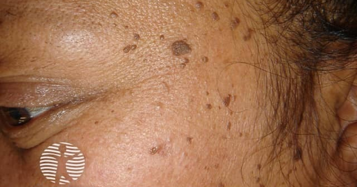
Dermatosis papulosa nigra is a common, entirely benign condition of Fitzpatrick IV–VI adults — multiple small, dark-brown to black, hyperpigmented papules on the malar cheeks, forehead and lateral neck. It is biologically a variant of seborrhoeic keratosis with strong familial clustering, typically appearing in adolescence and progressing through adulthood. Diagnosis is clinical. Treatment is cosmetic only; ablative modalities are highly effective but carry a substantial risk of post-inflammatory dyschromia in darker skin, so gentle techniques and conservative test patches are essential.
Clinical features
- Multiple small (1–5 mm), dark-brown to black, smooth or slightly verrucous papules.
- Distribution — malar cheeks, lateral forehead, temples, periorbital, lateral neck; less commonly upper chest and back.
- Affects Fitzpatrick IV–VI predominantly; up to 35% of Black adults in the UK and US population studies.
- Onset adolescence; numbers increase with age.
- Strong familial clustering — often multiple affected relatives.
- Asymptomatic.
Histology
- Identical to seborrhoeic keratosis — acanthotic epidermis with horn cysts and hyperpigmentation of the basal layer.
- Histology is rarely required for diagnosis.
Differential
- Verruca plana (flat warts) — smaller, smoother, in groups, often on dorsal hands.
- Acne / closed comedones — central white plug, periorbital and forehead.
- Cherry angioma — bright red, vascular.
- Acrochordon — pedunculated, neck.
- Pigmented BCC — single, slow-growing, atypical.
- Melanocytic naevi.
Management — and the dyschromia trap
- Reassurance and no treatment is the default — the condition is benign.
- Cosmetic options when patient requests:
- Snip or scissor excision (small papules).
- Light electrodesiccation or curettage.
- Cryotherapy — use with caution; high risk of post-inflammatory hypopigmentation in darker skin.
- Laser (KTP, pulsed dye, Er:YAG) — performed by clinicians experienced in skin of colour.
- Topical retinoids — mixed evidence.
- Always perform a test treatment on a single inconspicuous lesion before treating cosmetically prominent areas; counsel about the substantial risk of post-inflammatory hyper- or hypopigmentation.
- UV photoprotection complements treatment to limit new lesion development.
References
- Castellani A. Dermatosis papulosa nigra. Original description; 1925.
- Niang SO et al. Dermatosis papulosa nigra in Dakar, Senegal. Int J Dermatol; 2007.
- Taylor SC et al. Dermatology for skin of color. 2nd edition. McGraw-Hill; 2016.
Spot a correction?
If any clinical statement, citation or link on this page needs updating, please email admin@skinoncology.net with the page name, the proposed correction and the supporting source.