

Dermatofibroma
Benign fibrous histiocytoma; cutaneous fibrous histiocytoma; "BFH"; sclerosing haemangioma (older, mixed terminology)
The dermatofibroma is among the commonest benign cutaneous tumours and the single most frequent benign dermal nodule in adult skin clinics. Its clinical hallmark is a firm, well-demarcated, often pigmented dermal papule on the lower limbs that retracts beneath the skin surface on lateral compression — the "pinch / dimple sign". While the typical lesion is benign and unproblematic, several variants — cellular, aneurysmal, atypical, deep / plexiform — pose clinical and histological challenges and overlap with dermatofibrosarcoma protuberans (DFSP), atypical fibroxanthoma (AFX), pleomorphic dermal sarcoma (PDS) and dermal metastatic melanoma. CD34 immunohistochemistry — negative in dermatofibroma, positive in DFSP — is the key discriminator from the most clinically dangerous histological mimic. Treatment is unnecessary for typical lesions; excision with histology is appropriate for any clinically or histologically atypical case.
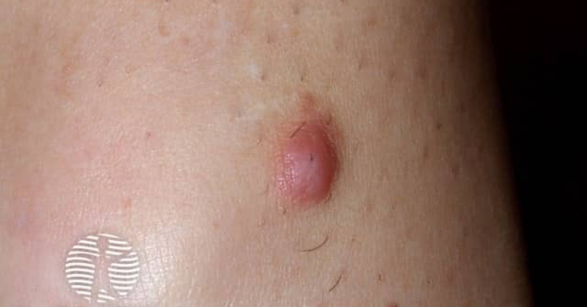
Clinical features
- Firm, well-demarcated, dome-shaped or slightly elevated dermal papule, 0.5–1.5 cm.
- Colour — flesh, pink, red-brown or dark brown; surface smooth or hyperpigmented.
- Pinch / dimple sign — lesion retracts beneath skin surface on lateral compression — pathognomonic.
- Distribution — lower limbs (especially shins of women) most common; also arms, trunk.
- Median age 20–50; F>M (~2:1).
- Often follows minor trauma (insect bite, ingrown hair) — historically considered a reactive process.
- Multiple dermatofibromas (>15) — consider associated immunosuppression (HIV, autoimmune disease, transplant).
Dermoscopy
- Central white scar-like patch ("white star", "white network") with a peripheral pigment network — the most characteristic dermatofibroma feature.
- Vascular pattern — fine or dotted vessels at the periphery of the central white area.
- Variants — homogeneous brown, multifocal pigmentation, atypical (when deep or aneurysmal).
Histology & variants
- Poorly circumscribed dermal proliferation of bland spindled fibroblasts and histiocytes in a storiform / cartwheel pattern.
- Overlying epidermis acanthotic with basal pigmentation ("dirty feet" appearance).
- Peripheral collagen trapping ("collagen cuffs") — characteristic.
- Immunohistochemistry — factor XIIIa+, CD68+, CD34− (the negative CD34 is the key distinction from DFSP, which is CD34+).
- Variants:
- Cellular dermatofibroma — markedly hypercellular; ~10% recurrence rate; rarely metastasises.
- Aneurysmal dermatofibroma — pseudovascular spaces with haemorrhage; can be confused with vascular tumour.
- Atypical "DF with monster cells" — bizarre nuclei in otherwise typical lesion.
- Deep / plexiform fibrohistiocytic tumour — extends into subcutis; intermediate behaviour with metastatic risk.
- Epithelioid fibrous histiocytoma — distinct entity, ALK-rearranged.
Critical differential — DFSP
- The most clinically important differential is dermatofibrosarcoma protuberans (DFSP) — a low-grade infiltrative dermal sarcoma that mimics a benign dermatofibroma clinically.
- Discriminating features:
- Clinically — DFSP is larger, more plaque-like, slowly enlarging over years, often on trunk / proximal limb (vs DF on distal lower limb).
- Histologically — DFSP is CD34+ and factor XIIIa− (opposite of dermatofibroma); infiltrative deep border into subcutis with a "honeycomb" pattern; uniform monotonous spindled cells.
- Molecularly — DFSP harbours COL1A1-PDGFB fusion.
- Other differentials — atypical fibroxanthoma (AFX), pleomorphic dermal sarcoma (PDS), dermal metastatic melanoma, leiomyosarcoma.
Management
- Reassurance — no treatment needed for typical, asymptomatic lesions.
- Excision with histology indicated for:
- Diagnostic uncertainty.
- Cosmetic concern.
- Recurrent symptoms (irritation, bleeding, snagging on clothing).
- Cellular, atypical, deep or plexiform variants.
- Multiple lesions in an immunocompromised patient (assess for occult disease).
- Margin — narrow (3–5 mm) excisional biopsy is sufficient for typical lesions.
- If histology suggests DFSP rather than DF: refer to skin cancer / sarcoma MDT for wide local excision (or Mohs micrographic surgery) and confirmatory COL1A1-PDGFB testing.
- Counsel that recurrence after complete excision is rare (5–10% in cellular variants, <5% in classical).
References
- Calonje E, Fletcher CD. Cutaneous fibrohistiocytic tumours. Histopathology; 2014.
- Han TY et al. Clinical and dermoscopic features of dermatofibroma. Ann Dermatol; 2011.
Spot a correction?
If any clinical statement, citation or link on this page needs updating, please email admin@skinoncology.net with the page name, the proposed correction and the supporting source.