

Cutaneous metastases
Secondary skin tumours; metastatic skin deposits
Cutaneous metastases occur in 0.7–9% of patients with internal malignancy. They may be the first manifestation of an undiagnosed cancer, herald disease recurrence, or signal advanced disease. The commonest primaries differ by sex: in women, breast carcinoma predominates, followed by melanoma, ovarian and lung; in men, classic series rank melanoma first, followed by lung and colorectal carcinoma. The clinical morphology often mimics benign processes — a firm dermal nodule, "rosacea-like" plaque or cellulitis-like erythema. Recognising and biopsying suspicious lesions is essential because management is determined by the primary tumour, and skin involvement carries prognostic significance.
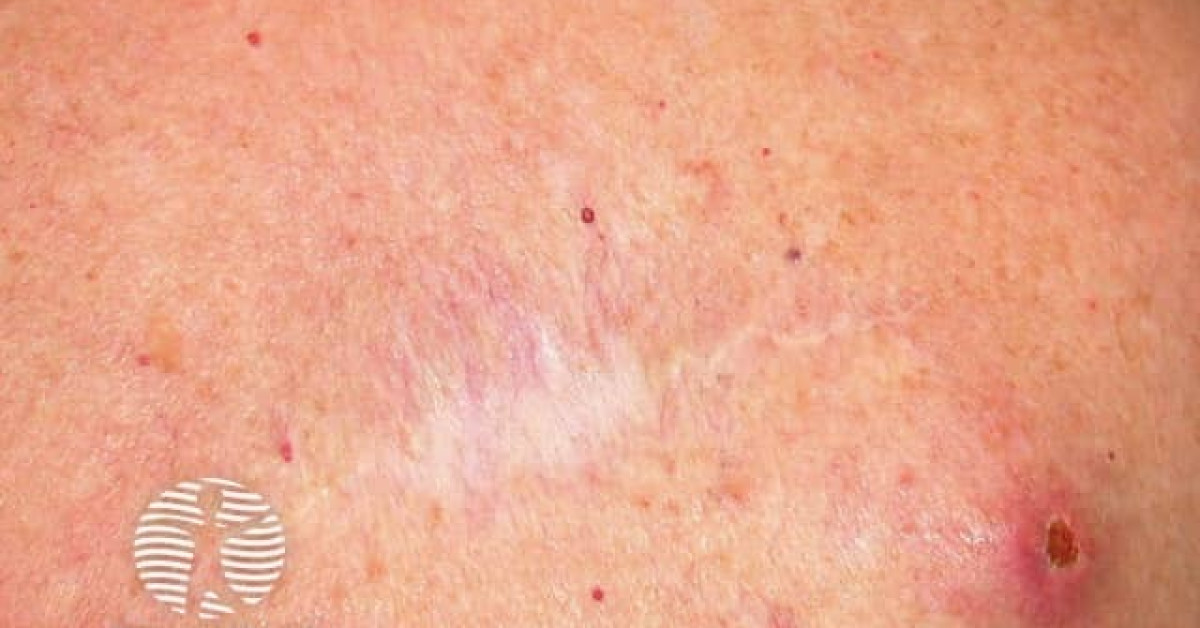
Epidemiology & commonest primaries
- Overall incidence in cancer patients: 0.7–9%; rises with stage IV disease.
- Women — breast carcinoma (~70% of cutaneous metastases in women); melanoma; ovarian; lung.
- Men — per Lookingbill 1993, melanoma is the commonest primary cutaneous metastasis (~32%), followed by lung carcinoma (~12%), colorectal (~11%); head & neck SCC and renal cell carcinoma also reported.
- Cutaneous deposits as the first sign of malignancy occur in 0.5–1% — most commonly lung and renal cell carcinomas.
Clinical patterns
- Solitary or multiple nodules — most common; firm, painless, dermal/subcutaneous, often mistaken for cysts or lipomas.
- Sister Mary Joseph nodule — umbilical metastasis; classically gastric, pancreatic, colorectal or ovarian primary; signifies poor prognosis.
- Carcinoma erysipeloides — well-demarcated red plaque mimicking erysipelas/cellulitis; classically breast cancer to chest wall via dermal lymphatics.
- Carcinoma en cuirasse — diffuse sclerotic infiltration of the chest wall; classical late breast carcinoma.
- Carcinoma telangiectoides — telangiectatic vascular plaques; breast and lung primaries.
- Alopecia neoplastica — scarring alopecic plaques on the scalp from breast and other primaries.
- Zosteriform metastases — dermatomal cutaneous deposits mimicking shingles.
Diagnosis
- Punch or excisional biopsy with adequate dermal/subcutaneous sample.
- Immunohistochemistry to characterise primary site:
- Breast — ER, PR, HER2, GATA3, mammaglobin.
- Lung — TTF-1, napsin A (adenocarcinoma); p40, p63 (squamous).
- Colorectal — CK20+, CDX2+, CK7−.
- Renal cell — PAX8+, RCC marker, vimentin.
- Ovarian — PAX8+, WT1+, CA-125.
- Prostate — PSA, NKX3.1.
- Melanoma — S100, SOX10, Melan-A, HMB-45.
- Imaging staging (CT chest/abdomen/pelvis ± PET-CT) when no known primary.
Management
- Primary disease driven — refer to or coordinate with the appropriate oncology team.
- Local options for symptomatic / single lesions:
- Excision for solitary, accessible, painful or bleeding lesions.
- Palliative external beam radiotherapy.
- Electrochemotherapy (NICE IPG446 for skin metastases from non-skin primary tumours and melanoma) — bleomycin + electroporation; useful for multiple superficial metastases especially breast (see monograph).
- Topical therapies (miltefosine, imiquimod) for superficial breast metastases.
- Intralesional chemotherapy or T-VEC (in melanoma).
- Systemic therapy of the primary tumour (chemotherapy, hormone therapy, targeted therapy, immunotherapy) addresses the underlying disease.
Prognosis
Cutaneous metastases generally indicate stage IV disease and a guarded prognosis — median survival 6–12 months, depending on primary tumour biology and response to systemic therapy. Some primaries (e.g. breast cancer with HER2-targeted therapy, melanoma with checkpoint inhibitors) have substantially better outcomes today than historically. Early biopsy of any suspicious cutaneous lesion in a known cancer patient — or a previously well patient with constitutional symptoms — is the most important diagnostic step.
References
- Wong CY et al. Cutaneous metastases: a review. Int J Dermatol; 2013.
- Lookingbill DP et al. Cutaneous metastases in patients with metastatic carcinoma. J Am Acad Dermatol; 1993.
Spot a correction?
If any clinical statement, citation or link on this page needs updating, please email admin@skinoncology.net with the page name, the proposed correction and the supporting source.