

Cutaneous mastocytosis
CM; urticaria pigmentosa (older — now "maculopapular cutaneous mastocytosis"); solitary mastocytoma; diffuse cutaneous mastocytosis; telangiectasia macularis eruptiva perstans (TMEP — older variant)
Cutaneous mastocytosis is a clonal proliferation of mast cells confined to the skin. The 2016 WHO classification recognises three principal clinical variants: maculopapular cutaneous mastocytosis (MPCM, formerly urticaria pigmentosa), the most common, with multiple yellow-tan to red-brown macules and papules; solitary mastocytoma, typically a single yellow-tan plaque on the trunk of an infant; and diffuse cutaneous mastocytosis, a rare severe pachydermatous infantile variant with widespread mast-cell infiltration. Childhood-onset disease (~80% of cases) is generally indolent and frequently resolves by adolescence; adult-onset cutaneous mastocytosis is rare and very strongly associated with underlying systemic mastocytosis — most often indolent systemic mastocytosis, but with a meaningful minority progressing to advanced systemic disease (smouldering, aggressive, mast-cell leukaemia or mast-cell sarcoma). The somatic KIT D816V mutation drives the great majority of disease. The clinical hallmark — the Darier sign (urtication on stroking a lesion) — is pathognomonic.
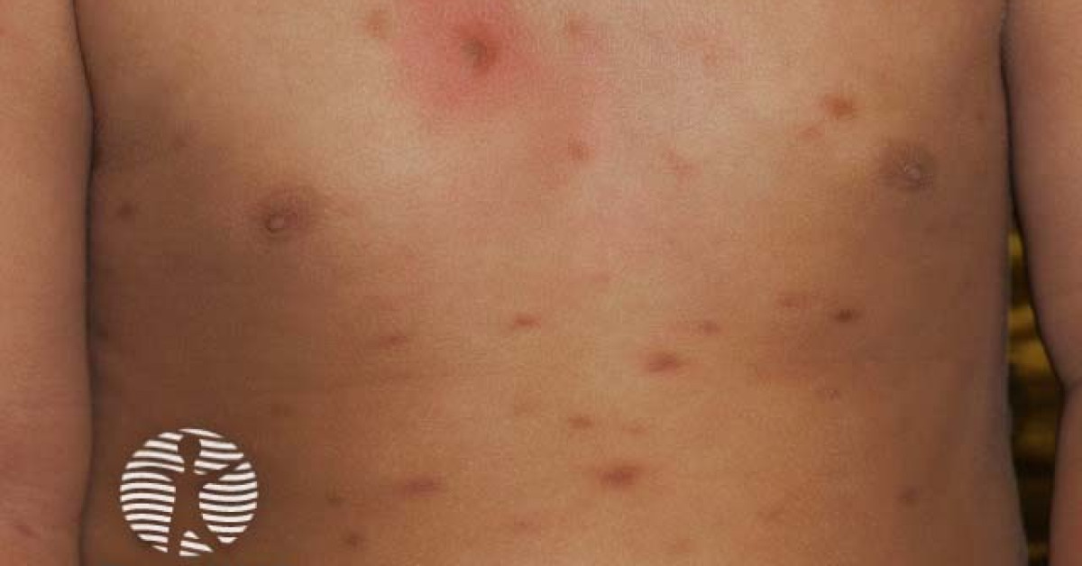
Variants
- Maculopapular cutaneous mastocytosis (MPCM) — formerly urticaria pigmentosa; the commonest variant. Multiple yellow-tan to red-brown macules and papules concentrated on the trunk; in children spares face, palms and soles, in adults can involve any site. Polymorphic variant (children) — variable lesion size, irregular shape; usually resolves by adolescence. Monomorphic variant (adults) — uniformly small monomorphic lesions, persists, often associated with systemic mastocytosis.
- Solitary mastocytoma — single yellow-tan to red-brown plaque, 1–4 cm, on the trunk of an infant. Strongly Darier-sign-positive. Usually resolves by adolescence.
- Diffuse cutaneous mastocytosis — widespread thickened "leather-like" infiltration of skin in infants. Severe; risk of life-threatening mast-cell mediator release (anaphylaxis, hypotension, GI symptoms).
- Telangiectasia macularis eruptiva perstans (TMEP) — older variant; subtle telangiectatic patches with fewer mast cells; often progresses to systemic disease in adults.
Clinical features & mediator release
- Darier sign — urtication / wheal-and-flare reaction on stroking a lesion (mast-cell degranulation) — pathognomonic.
- Triggers — heat, friction, NSAIDs, opiates, alcohol, contrast media, anaesthetic agents (atracurium, succinylcholine), insect stings, exercise.
- Mediator-release symptoms — pruritus, flushing, urticaria, headache, abdominal pain, diarrhoea, palpitations, syncope, anaphylaxis.
- Severe / life-threatening anaphylaxis can occur, particularly in diffuse cutaneous and aggressive systemic disease.
- Childhood-onset — generally indolent, often resolves by adolescence; rarely systemic.
- Adult-onset — strongly associated with systemic mastocytosis (~80% have systemic disease at presentation or develop it during follow-up); persistent throughout life.
Histology & molecular
- Increased mast cells in the upper / mid dermis (sometimes throughout the dermis in diffuse variants).
- Mast cells highlighted by toluidine blue (metachromatic), CD117 (KIT) and tryptase IHC.
- Aberrant mast-cell phenotype in mastocytosis (vs reactive) — aberrant CD25 expression on mast cells is highly suggestive.
- KIT D816V mutation in >90% of adult mastocytosis (sensitive PCR or NGS); less commonly in childhood disease.
- Tryptase staining helps quantify mast-cell density (>15 / HPF in true mastocytosis).
Workup for systemic disease
- Particularly important in adults with new cutaneous mastocytosis — exclude / confirm systemic involvement.
- Serum tryptase — >20 ng/mL is one of the WHO minor criteria for systemic mastocytosis (per WHO-HAEM5, interpret in the context of hereditary alpha-tryptasemia / TPSAB1 copy number, which raises baseline tryptase); persistent elevation warrants bone marrow.
- Bone marrow biopsy with KIT D816V testing — diagnostic gold standard; aberrant mast-cell aggregates (>15 cells), spindled morphology, aberrant CD25 expression, KIT D816V mutation are the WHO criteria.
- FBC, LDH, alkaline phosphatase, B-type symptom assessment.
- DEXA scan — osteoporosis common in systemic mastocytosis.
- Refer to a specialist mastocytosis service (UK: King's College London / Royal Marsden / specialist allergy services).
Management
- Trigger avoidance — patient education about heat, friction, alcohol, NSAIDs, opiates, certain drugs and procedures (anaesthesia consultation pre-operatively).
- Symptomatic mediator-release management:
- H1 antihistamines (cetirizine, loratadine, fexofenadine) — first-line.
- H2 antihistamines (famotidine) — for GI symptoms.
- Mast-cell stabilisers (cromolyn sodium oral / topical).
- Leukotriene receptor antagonists (montelukast).
- Topical corticosteroids for symptomatic skin lesions.
- PUVA / narrowband UVB phototherapy for symptomatic widespread disease.
- Anaphylaxis prevention — adrenaline auto-injector (two devices); medic-alert; written emergency action plan; venom immunotherapy if Hymenoptera trigger.
- Advanced systemic disease — KIT-targeted therapy (midostaurin, avapritinib, ripretinib), interferon-α, cladribine, allogeneic transplantation in selected cases (managed by specialist mastocytosis service).
- Solitary mastocytoma — observation; surgical excision rarely needed.
- Childhood mastocytosis — most resolve by adolescence; reassurance plus trigger avoidance.
References
- Valent P et al. Updated diagnostic criteria and classification of mast cell disorders — a consensus proposal. HemaSphere; 2021.
- Hartmann K et al. Cutaneous manifestations in patients with mastocytosis — consensus report. J Allergy Clin Immunol; 2016.
Spot a correction?
If any clinical statement, citation or link on this page needs updating, please email admin@skinoncology.net with the page name, the proposed correction and the supporting source.