

Bowenoid papulosis
Multifocal HPV-driven anogenital intraepithelial neoplasia in young adults
Bowenoid papulosis is a clinico-pathological entity in which multiple small, pigmented or red papules arise on the anogenital skin of young, otherwise healthy adults, driven by high-risk human papillomavirus (most commonly HPV-16). The defining feature is a striking discordance between an alarming histological appearance — full-thickness keratinocyte atypia indistinguishable from squamous cell carcinoma in situ (Bowen's disease) — and a generally benign clinical course, with frequent spontaneous regression and a low rate of progression to invasive carcinoma. Recognition matters because aggressive surgical management of every lesion is unnecessary; topical or destructive therapy with surveillance is usually appropriate. Sexual partner screening, cervical cytology, HIV screening and HPV vaccination should be considered.
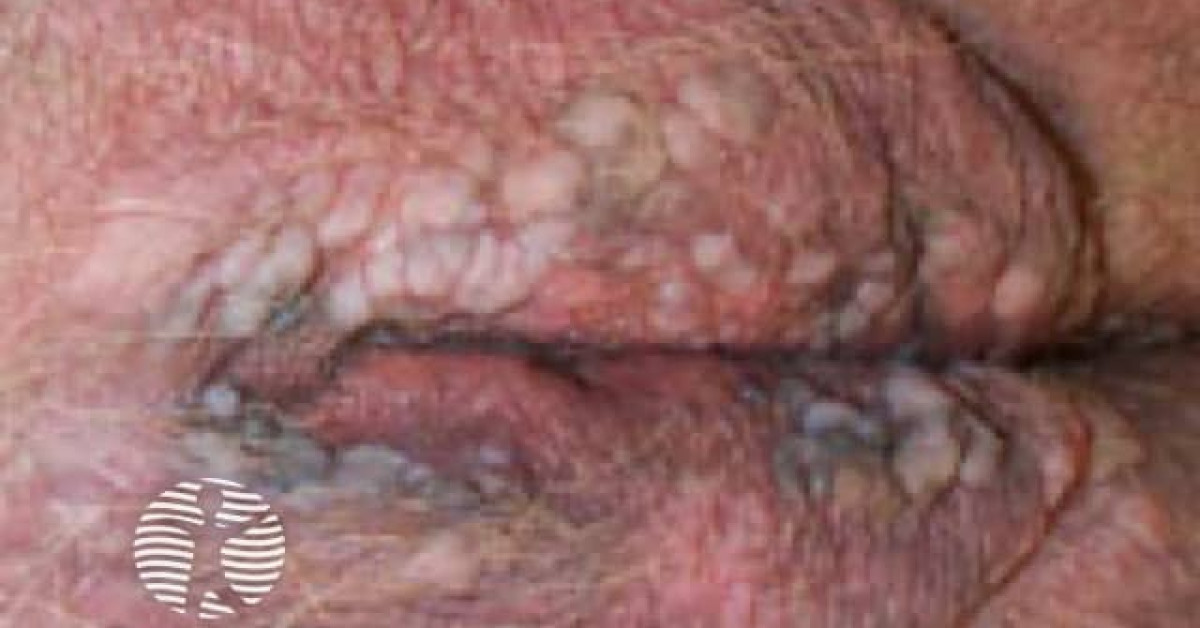
Clinical features
- Multiple (often 4 to 30+) small (2–10 mm) pigmented brown to violaceous papules with a smooth or velvety surface.
- Distribution: penile shaft and glans (men); vulva, perianal area (women); perianal in both sexes.
- Median age 25–35; both sexes; sexually active patients.
- Usually asymptomatic; occasional itch or soreness.
- Frequently misdiagnosed as warts (condyloma acuminatum), seborrhoeic keratoses, naevi or melanoma.
Differential diagnosis
- Anogenital warts (condyloma acuminatum) — usually verrucous, skin-coloured to white, less pigmented, low-risk HPV (6, 11).
- Vulvar / penile intraepithelial neoplasia (VIN, PeIN) — usually fewer, larger, less pigmented lesions; behaves more aggressively. The histological line between bowenoid papulosis and HPV-driven uVIN/PeIN is blurred — the distinction is largely clinical (multifocal small papules in a young patient = bowenoid papulosis; large solitary plaque in older patient = VIN/PeIN).
- Erythroplasia of Queyrat — solitary or few large red velvety plaque on glans of older uncircumcised man.
- Bowen's disease — solitary scaly plaque on sun-exposed or anogenital skin of older adult.
- Pigmented seborrhoeic keratosis, melanocytic naevus — clinical mimics.
Diagnosis
- Punch biopsy of representative lesion(s).
- Histology indistinguishable from Bowen's disease — full-thickness keratinocyte atypia with mitoses at all levels — but in a multifocal anogenital field of a young adult.
- p16 strongly diffuse positive (HPV-driven); high-risk HPV typing (especially HPV-16) supports diagnosis.
- Examine the entire anogenital area, oral mucosa and assess for cervical disease in women.
- HIV testing in selected patients.
Management
- Conservative — many lesions regress spontaneously over months to years, particularly in young patients with intact immunity.
- Topical 5% imiquimod (3 nights/week for 12–16 weeks) — clearance in 50–80%.
- Topical 5-fluorouracil — alternative.
- Cryotherapy, curettage, electrocautery, CO₂ laser — for selected larger/persistent lesions.
- Surgical excision reserved for atypical, suspicious or refractory lesions.
- Counsel about sexual transmission, condom use and partner examination.
- Cervical cytology and HPV screening for the patient and female partners.
- HPV vaccination — primary prevention for partners and unaffected individuals; may reduce recurrence in treated patients.
Prognosis
Excellent — most cases regress or are cleared with conservative treatment. Progression to invasive squamous cell carcinoma is uncommon in young immunocompetent patients (<5% in older cohorts); modern series including older patients and HIV-positive or otherwise immunocompromised cohorts report progression rates of 10–15%. Long-term surveillance (annual review and prompt biopsy of any persistent or atypical lesion) is appropriate, particularly in immunocompromised patients.
References
- Wade TR et al. Bowenoid papulosis of the genitalia. Arch Dermatol; 1979.
- Schwartz RA, Janniger CK. Bowenoid papulosis. J Am Acad Dermatol; 1991.
Spot a correction?
If any clinical statement, citation or link on this page needs updating, please email admin@skinoncology.net with the page name, the proposed correction and the supporting source.