Blue naevus & cellular blue naevus
Common (Jadassohn-Tièche) blue naevus; cellular blue naevus (CBN); atypical cellular blue naevus (ACBN); malignant blue naevus (rare)
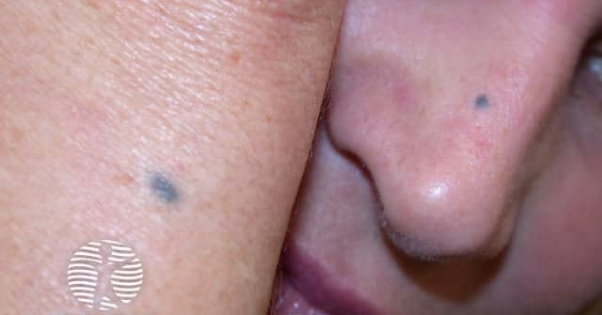
Blue naevi are benign dermal melanocytic proliferations composed of pigmented dendritic and spindled melanocytes deep in the dermis. The deep position of melanin gives the characteristic blue-grey colour by the Tyndall effect. Common blue naevi are small, stable and benign. Cellular blue naevi are larger, sometimes worrisome variants that — particularly in the atypical form — may mimic melanoma both clinically and histologically. Driver mutations in GNAQ and GNA11 overlap those of uveal melanoma. Malignant blue naevus (melanoma arising in or resembling a cellular blue naevus) is exceptionally rare but aggressive, and follows the molecular path typical of GNAQ/GNA11-mutant melanomas including BAP1 loss as a marker of progression.
Subtypes
- Common (Jadassohn-Tièche) blue naevus — small (<1 cm), well-circumscribed, slate-blue macule or papule; dorsal hand, foot, scalp; usually present from childhood / adolescence; benign.
- Cellular blue naevus (CBN) — larger (1–3 cm), nodular, blue-black; buttock, sacrum, scalp; histologically more cellular with biphasic architecture.
- Atypical cellular blue naevus (ACBN) — borderline lesion with atypical features (mitoses, atypia, necrosis); behaviour uncertain.
- Malignant blue naevus / melanoma ex blue naevus — exceptionally rare; histologically a melanoma arising within or resembling a CBN; aggressive metastatic potential.
- Special variants: epithelioid blue naevus (Carney complex association), deep penetrating naevus (overlapping spectrum).
Clinical features
- Slate-blue, blue-grey or blue-black macule, papule or nodule.
- Common blue naevus typically <1 cm; cellular blue naevus 1–3 cm or larger.
- Predilection sites: dorsa of hands and feet (common); buttock and sacrum (cellular).
- Stable for years; rapid change, ulceration or sudden growth warrants excision.
- Differential: nodular melanoma (especially amelanotic with focal pigment), Spitz naevus (pigmented variant), dermatofibroma with haemosiderin, glomus tumour, traumatic tattoo.
Dermoscopy
- Steel-blue or homogeneous blue-grey pattern (no network, no globules, no streaks).
- Symmetrical pigmentation favours benign blue naevus.
- Asymmetry, multicomponent pattern, peripheral structures, ulceration favour cellular variants or melanoma.
Histology & molecular
- Common blue naevus — pigmented dendritic and spindled melanocytes scattered in the reticular dermis with abundant melanophages; HMB-45+, S100+.
- Cellular blue naevus — biphasic: pigmented dendritic component plus densely cellular nodules of plump spindled cells; "dumbbell" extension into subcutis common.
- ACBN — atypia, mitoses, focal necrosis or expansive growth raise concern; Ki-67 elevated.
- Driver mutations: GNAQ or GNA11 (~85%) — same as in uveal melanoma; rarely CYSLTR2 / PLCB4.
- BAP1 loss by IHC is a marker of progression toward malignancy in atypical blue naevi.
Management
- Stable, classical common blue naevi do not require excision — observation with photographic surveillance is appropriate.
- Excisional biopsy with complete histology for any:
- Atypical, large or rapidly changing blue lesion.
- Cellular blue naevus, particularly >1 cm or with atypical features.
- Lesion in an unusual site (e.g. trunk, head/neck of an adult) or arising de novo in adulthood.
- For atypical cellular blue naevus / probable malignant blue naevus — wide local excision with margins per Breslow thickness if invasive melanoma is confirmed; sentinel lymph node biopsy by AJCC criteria.
- Molecular profiling (GNAQ/GNA11 + BAP1) helpful in equivocal cases.
- Genetic counselling for BAP1-loss tumours — see BAP1-TPDS.
References
- Murali R et al. Blue naevi and related lesions — a review of clinical, histological and molecular features. Pathology; 2009.
- Van Raamsdonk CD et al. Frequent somatic mutations of GNAQ in uveal melanoma and blue naevi. Nature; 2009.
Spot a correction?
If any clinical statement, citation or link on this page needs updating, please email admin@skinoncology.net with the page name, the proposed correction and the supporting source.